
Abstract
Cerebral autosomal dominant arteriopathy with subcortical infarcts and leukoencephalopathy (CADASIL), the most common monogenic small vessel disease, is caused by cysteine-altering mutations in the neurogenic locus notch homolog protein 3 gene, which disrupt vascular smooth muscle cell function through protein misfolding and impaired vascular reactivity. Neuroimaging, particularly high-resolution magnetic resonance imaging, reveals characteristic white matter hyperintensities, and advanced imaging combined with deep learning enhances diagnostic precision. Blood neurofilament light chain is an emerging biomarker. Genetic counselling is crucial for early diagnosis and family screening. Current management involves vascular risk reduction, migraine control, and supportive care. Investigational therapies, including monoclonal antibodies, exon-skipping antisense oligonucleotides, CRISPR-based gene editing, and ongoing trials such as AMCAD and tocotrienol (NCT04658823), hold promise. Challenges remain in biomarker validation, therapeutic translation, and equitable access to diagnostics, highlighting the need for international collaboration to optimise CADASIL care.
Keywords: CADASIL, NOTCH3 mutation, biomarkers, neuroimaging, vascular dementia, small vessel disease
Introduction
Cerebral autosomal dominant arteriopathy with subcortical infarcts and leukoencephalopathy (CADASIL) is the most prevalent monogenic small vessel disease, caused by heterozygous cysteine-altering variants in the neurogenic locus notch homolog protein 3 (NOTCH3) extracellular domains NOTCH3 gene (). These mutations result in progressive cerebral small-vessel pathology, ultimately causing ischaemic strokes, leukoencephalopathy, and vascular dementia (). Advances in whole-exome and genome sequencing have enabled genotype-phenotype correlations, notably linking Epidermal growth factor-like repeat (EGFr) domains 1-6 with early stroke onset and poor outcomes (). Immunotherapies targeting aggregated NOTCH3 extracellular domains (NOTCH3ECD) have shown preclinical efficacy (). High-resolution magnetic resonance imaging (MRI) enhances lesion characterisation, particularly white matter hyperintensities (WMHs) in the anterior temporal lobes and external capsules, which aid in diagnosis and tracking disease progression (,). Blood neurofilament light chain (NfL) is an emerging non-invasive biomarker correlating with imaging and clinical severity. This review highlights CADASIL’s genetic basis, advances in sequencing, neuroimaging, and management, while underscoring emerging priorities: risk prediction, circulating/imaging biomarkers, NOTCH3ECD-dependent WMHs-and the role of international collaborative networks in translational research and clinical care ().
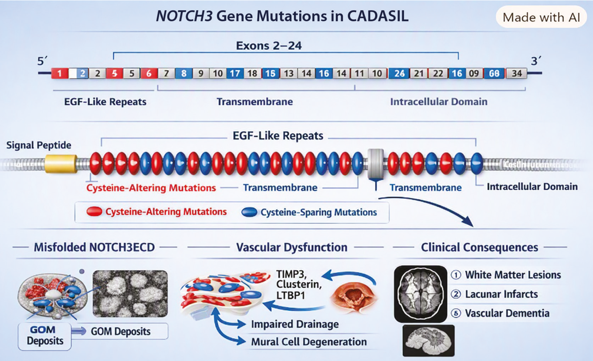
Schematic representation of the NOTCH3 gene and cysteine dependent mutations in CADASIL. The NOTCH3 gene comprises 33 exons encoding multiple domains, with most pathogenic variants located within the epidermal growth factor (EGF) like repeats (exons 2–24). Cysteine altering mutations (red) disrupt disulphide bonding, leading to misfolded NOTCH3 extracellular domain, granular osmiophilic material (GOM) deposition, and vascular dysfunction. Cysteine sparing mutations (blue) are less pathogenic. The schematic illustrates the relationship between molecular alterations, impaired extracellular matrix organization, and clinical consequences including white matter lesions and vascular dementia ()
Literature Search Strategy
This narrative review was based on a structured search of PubMed/MEDLINE, Scopus, Web of Science, and Google Scholar for studies published between January 2000 and March 2026. Keywords included “CADASIL”, “NOTCH3”, “genetics”, “biomarkers”, “MRI”, “neuroimaging”,
“gene-therapy”, “CRISPR”, “vascular dementia”, and “small vessel disease”. The search identified about 1,240 records; after removing duplicates, 980 were screened by title and abstract. Of these, 660 full texts were assessed, and 410 were excluded for irrelevance, non-English language, or lack of full text. A total of 250 studies were included, comprising original research, systematic reviews, meta-analyses, clinical trials, consensus statements, and clinical guidelines. summarises selected CADASIL studies by study design, population or experimental model, and key findings.
Table 1. Selected studies and recommendations on the epidemiology, genetics, diagnosis, biomarkers, and management of CADASIL
Author | Year | Study type | Sample/model | Key findings |
|---|---|---|---|---|
Yamamoto et al. () | 2023 | Review | NA | Updated epidemiology, biomarkers, and pathogenesis of CADASIL |
Choi () | 2010 | Review | NA | Genetic basis of CADASIL and NOTCH3 mutations |
Oliveira et al. () | 2022 | Experimental | Mouse model | Immunotherapy reduces NOTCH3 aggregation |
Rutten et al. () | 2020 | Cohort | UK Biobank (>1000) | High prevalence and broad phenotype of NOTCH3 variants |
Kim et al. () | 2022 | Observational | Asian cohort | Ethnic differences in phenotype and imaging features |
Gravesteijn et al. () | 2025 | Cohort | 1900+ | Disease severity staging system (NOTCH3-SVD) |
Anisetti et al. () | 2023 | Cross-sectional | Multicenter | CADASIL clinical grading system validation |
Alirezaei et al. () | 2019 | Review | NA | NfL correlation with MRI and disease severity |
Meschia et al. () | 2023 | Guideline | AHA statement | Management recommendations for CADASIL |
CADASIL: Cerebral autosomal dominant arteriopathy with subcortical infarcts and leukoencephalopathy, NOTCH3: Neurogenic locus notch homolog protein 3, MRI: Magnetic resonance imaging
Epidemiology and Natural History
Prevalence and Demographic Distribution
Genomic analyses using large databases such as ExAC and UK Biobank have significantly revised CADASIL prevalence estimates, identifying pathogenic NOTCH3 variants in up to one in three hundred individualssubstantially higher than earlier registry-based estimates () (). Regional variation is evident, with the p.Arg544Cys variant found in 0.85% of the Taiwanese population. Demographic factors influence clinical presentation. Asian patients tend to have a later symptom onset (mid-50s) and a lower migraine prevalence (2.5-33% vs. 40-54%) compared to Western cohorts (mid-30s). Intracerebral haemorrhage and cerebral microbleeds are more common in Asian patients (25% and 87.5%, respectively) than in European patients (<1% and 31%, respectively). Conversely, anterior temporal pole WMHs-a hallmark feature-are less frequently observed in Asians (20-44.5%) than in Europeans (89-93%) ().
Disease Progression and Clinical Milestones
CADASIL severity is measured by the NOTCH3-SVD Staging System, integrating Fazekas’ scores, lacune count, and mRS, with 94% showing sequential progression (), and the Clinical CADASIL Grading System, demonstrating high inter-rater reliability (κ=0.855) (). Long-term studies reveal 85% progression over 18 years, correlating more strongly with imaging and biological markers-lacune burden, parenchymal loss, and NOTCH3 aggregation-than with clinical status, age, or sex (,,).
Genetic Basis of CADASIL and Clinical Genetic Counselling Guidelines
CADASIL arises from cysteine-altering mutations within the 34 EGFr repeats of the NOTCH3 gene (). These disrupt disulphide bonding, causing extracellular domain misfolding, aberrant bridge formation, aggregation, and ER retention. Misfolded NOTCH3ECD accumulates as granular osmiophilic material (GOM) in VSMCs and pericytes, sequestering TIMP3, clusterin, and LTBP-1, disrupting perivascular homeostasis (,). This impairs cerebrovascular reactivity and drainage and promotes degeneration, leading to hypoperfusion, white-matter lesions, and vascular dementia (). Pathogenic variants include P.R544C and p.Arg133Cys, with EGFr 7-34 linked to broader small vessel disease phenotypes (-).
Genetic Testing Indications
Genetic testing for NOTCH3 is recommended when clinical, imaging, and familial features suggest CADASIL, including early-onset (<55 years) ischemic events, recurrent migraine with aura (), confluent leukoencephalopathy in anterior temporal poles/external capsules, and a family history of stroke or cognitive decline. Confirmation requires identifying cysteine-altering EGFr variants via DNA sequencing (). VUS are assessed using ACMG/AMP criteria (PS4, PP4, PM2, PS3) (), refined by ClinGen guidelines emphasising cysteine-altering EGFr variants (), with further resolution through segregation, computational, and domain-based stratification.
Clinical manifestations and MRI features of CADASIL, showing common symptoms (small subcortical infarcts, migraines with aura, cognitive impairment, psychiatric disturbances) and hallmark imaging findings (white matter hyperintensities, lacunes, cerebral microbleeds, enlarged perivascular spaces) in characteristic brain regions ()
Biomarkers and Molecular Pathways
Emerging Molecular Pathways
Proteomic analyses in CADASIL vasculature reveal impaired ECM organisation, disrupted VSMC cytoskeletal dynamics, and defective proteostasis, promoting aggregation of mutant NOTCH3ECD (). This drives autophagic dysfunction, dysregulated angiogenesis, and TGF-β signalling, leading to fibrotic microvascular remodelling (). Mechanistically, N3ECD accumulation induces oxidative stress via Nox5, activating ER stress and RhoA/Rho kinase pathways, while disrupted calcium homeostasis impairs eNOS-sGC-cGMP signalling, compromising endothelial integrity (,). Targeted therapies-antisense oligonucleotide (ASO) for exon skipping, monoclonal antibodies against NOTCH3ECD, RNA interference, and CRISPR-mediated editing-show promise in reducing aggregates, restoring vascular function, and correcting pathogenic alleles in preclinical models ().
Fluid and Imaging Biomarkers
Neurofilament light chain in blood is a validated biomarker of neuroaxonal injury in CADASIL. Elevated levels correlate with white matter lesion burden, cognitive decline, and long-term disability and predict outcomes over 7-17 years, supporting its use in disease monitoring. In contrast, although microRNAs have been studied in related cerebrovascular disorders, they have not been clinically validated for CADASIL. Retinal imaging detects early microvascular changes in CADASIL, manifested as reduced vascular density on optical coherence tomography angiography (OCTA) and an increased wall-to-lumen ratio on adaptive optics. However, these alterations do not correlate with MRI-defined lesion burden or clinical severity, limiting their prognostic and diagnostic applicability (,).
Challenges in Ensuring Consistent and Reproducible Results for Biomarker Measurement
The reproducibility of biomarker findings in CADASIL is limited by small cohort sizes, single-centre designs, and methodological inconsistencies in sample handling, assay platforms, and analytical protocols. While high-throughput techniques like mass spectrometry and multiplex immunoassays hold potential, they require rigorous standardisation and transparent performance reporting to be clinically useful (). Multicentre collaborations with harmonised methodologies and open data sharing are essential for advancing biomarker validation in CADASIL.
Advances in Diagnostic Modalities
MRI and Advanced Neuroimaging
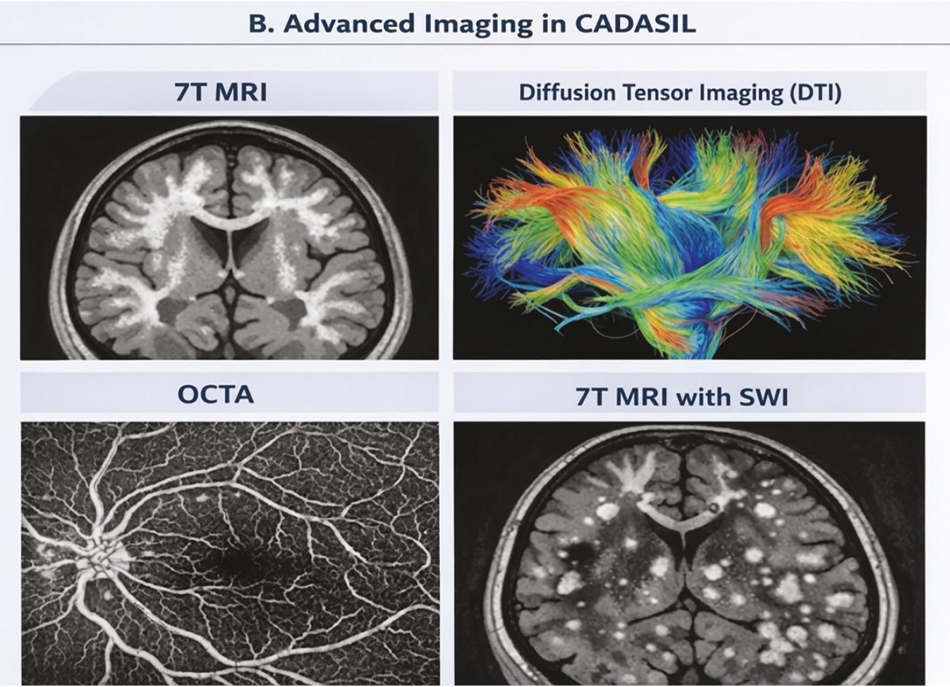
In CADASIL, hallmark WMHs occur bilaterally in anterior temporal poles and external capsules, with high diagnostic specificity even in early stages (,,). WMHs also show symmetrical periventricular and deep white matter distribution, sometimes extending into the corpus callosum. Lacunar infarcts in basal ganglia, thalamus, centrum semiovale, and pons correlate with cognitive decline (,).
Temporal pole WMHs differ pathologically, reflecting enlarged perivascular spaces and myelin loss (). Quantitative tools-CADA-MRIT and CADASILSeverity Grading System-standardise lesion assessment, enhance diagnostic accuracy and improve trial stratification () ().
Limitations of Conventional MRI in CADASIL
Despite its diagnostic utility, conventional MRI has limited prognostic sensitivity in CADASIL; imaging markers such as WMHs and lacunes correlate poorly with clinical severity and do not reliably predict disease progression. Furthermore, standard MRI lacks the spatial resolution necessary to evaluate microvascular hemodynamics or to detect early perfusion changes, limiting its capacity to capture preclinical disease activity ().
Quantitative and High-Field Techniques
Quantitative neuroimaging enhances CADASIL assessment. SIENA-derived PBVC shows ~0.45% annual atrophy, with incident lacunes predicting decline better than WMH progression (). Diffusion tensor imaging reveals increased mean diffusivity and reduced peak height in normal-appearing white matter, enabling early detection (). Ultra-high-field 7T SWI visualises microbleeds and vascular changes, including the “Chocolate Chip Sign” and 14.6% venous density reduction (). Deep learning CNNs improve lesion segmentation for WMHs, lacunes, microbleeds, and perivascular spaces (), with Dice ~0.75-0.85 (). Limitations include high cost, infrastructure demands, implant incompatibility, methodological variability, and lack of clinician training (,).
Retinal and Extracerebral Imaging Windows
Retinal vascular changes-arterial narrowing, arteriovenous nicking, and reduced OCTA vessel density-may indicate early CADASIL pathology but correlate poorly with symptoms or MRI lesion burden (,,). Reliability is limited by artefacts, segmentation errors, and protocol variability, especially in OCTA (). Standardised acquisition, quality control, and analysis protocols are urgently needed for research and clinical use. compares conventional MRI, diffusion tensor imaging, 7-T MRI, and optical coherence tomography angiography in terms of what each technique detects and its main limitations.
Table 2. Advantages and limitations of imaging modalities used in CADASIL
Imaging Method | Advantage | Limitation |
Conventional MRI | High sensitivity for lesion detection | Poor prognostic value |
Diffusion tensor imaging (DTI) | Detects microstructural changes | Protocol variability |
7 Tesla MRI (7T MRI) | High-resolution vascular imaging | Expensive and limited availability |
Optical coherence tomography angiography (OCTA) | Early microvascular detection | Poor correlation with clinical severity |
MRI: Magnetic resonance imaging
Laboratory Biomarkers
Genetic testing for pathogenic NOTCH3 mutations remains the gold-standard assay for CADASIL, performed on blood DNA (). No protein- or metabolite-based biomarker has achieved validation; CSF amyloid-β42, total tau, and phosphorylated tau lack specificity and are not recommended. Neurofilament light chain in blood and CSF correlates with severity, lesion burden, cognition, and outcomes, supporting monitoring utility (,). Emerging biomarkers include CNS-derived extracellular vesicles (EVs) carrying disease-specific cargo (), though clinical use is limited by isolation variability, purity, and specificity; omics-based EV profiling is under investigation ().
Current Management Strategies
General Principles of Care
Effective management of CADASIL centres on intensive blood pressure control and smoking cessation-interventions strongly associated with reduced risk of stroke and cognitive decline. Although evidence for treating comorbid dyslipidemia remains limited, lifestyle modifications and comprehensive vascular risk control contribute to slowing disease progression (). Pharmacologic therapies, particularly antiplatelet and anticoagulant agents, require individualised risk-benefit assessments due to the elevated risk of intracerebral haemorrhage; routine use of antiplatelet agents is not recommended in patients without prior ischemic events (). summarises clinical recommendations for confirming the diagnosis, counselling at-risk families, monitoring disease progression, and managing vascular risk and symptoms.
Table 3. Clinical recommendations for the diagnosis, management, and genetic counselling of patients with CADASIL
Clinical domain | Current recommendation |
|---|---|
Blood pressure management | Maintain blood pressure <130/80 mmHg through intensive vascular risk control |
Smoking cessation | Complete smoking cessation is strongly recommended |
Genetic diagnosis | Confirm diagnosis by identifying pathogenic NOTCH3 mutations using genetic testing |
Genetic counselling | Offer pre-test and post-test genetic counselling and cascade testing for at-risk relatives |
Neuroimaging | Brain MRI is recommended for diagnosis and monitoring disease progression |
Antiplatelet therapy | Consider after ischemic stroke or TIA; not routinely recommended for primary prevention |
Anticoagulation | Use only for standard indications (e.g., atrial fibrillation) after individualized risk-benefit assessment |
Symptomatic management | Appropriate treatment of migraine, cognitive impairment, psychiatric symptoms, and rehabilitation should be individualized |
NOTCH3: Neurogenic locus notch homolog protein 3, MRI: Magnetic resonance imaging
Antithrombotic and Anticoagulant Therapy
Evidence on antithrombotic therapy in CADASIL is limited. In a TriNetX cohort (n=455), antiplatelet use did not alter ischemic stroke or ICH risk (). A 2024 review of 142 ICH cases found anticoagulants-not antiplatelets-associated with higher hemorrhagic risk (20.0% vs. 1.9%, p=0.006), with ICH in 5.9% overall and 6.7% linked to anticoagulants (,). Low-dose aspirin after ischemic events appears generally safe (), though primary prevention remains uncertain. Anticoagulation is reserved for atrial fibrillation (CHA2DS2-VASc ≥2) or venous thromboembolism, with alternatives like left atrial appendage occlusion considered in patients with extensive microbleeds.
Acute Reperfusion Therapies
Evidence for acute reperfusion in CADASIL is limited. Individualised intravenous thrombolysis with alteplase has shown favourable outcomes; in a multicentre series (n=12), 83% achieved mRS 0-1 at 90 days without symptomatic haemorrhage (,). Consensus discourages thrombolysis for lacunar strokes but supports cautious use in large-vessel occlusions, contingent on MRI microbleed absence and close monitoring. Mechanical thrombectomy has shown promise in rare large-vessel occlusions, with successful reperfusion yielding clinical improvement without haemorrhagic complications ().
Symptomatic and Supportive Treatments
Migraine Management in CADASIL
Acute migraine in CADASIL is managed with analgesics (paracetamol, ibuprofen, aspirin) and abortive agents like sumatriptan, which-despite vasoconstrictive concerns-has not been clearly linked to ischemic events and is considered safe short-term with individualised risk assessment (,). Prophylaxis includes amitriptyline, pizotifen, acetazolamide, and sodium valproate, while propranolol is avoided due to adverse effects (,). Emerging CGRP-targeted therapies (gepants, ditans) may be less vasoconstrictive but require caution until CADASIL safety is established (,).
Cognitive, Psychiatric, and Rehabilitative Therapies
Evidence supporting targeted interventions for neuropsychiatric symptoms in CADASIL remains sparse, with most treatment regimens adapted from general psychiatric practice rather than CADASIL-specific trials. The American Heart Association notes that optimal strategies for these symptoms have yet to be clearly defined. Nonetheless, selective serotonin reuptake inhibitors are frequently prescribed in clinical settings, although their efficacy in CADASIL has not been systematically validated ().
Pharmacological Management of Neuropsychiatric Symptoms
Depression
Selective serotonin reuptake inhibitors are routinely used and generally well tolerated in treating depressive symptoms in CADASIL, with dose escalation considered for suboptimal responders. Although amitriptyline has demonstrated limited benefit, its use is discouraged in patients with cognitive impairment because of its anticholinergic effects. Sodium valproate may theoretically exert benefits via modulation of NOTCH3 receptor activity; however, mood disturbances in CADASIL are frequently resistant to pharmacotherapy. In rare, treatment-refractory melancholic cases, electroconvulsive therapy has been employed as a last-line intervention ().
Psychosis
Short-term administration of antipsychotics, particularly atypical agents, has been used to manage psychotic symptoms such as agitation, aggression, and excitatory states. In one reported case, a combination of lithium and a high-potency typical antipsychotic was effective in treating mixed manic symptoms ().
Cognitive Dysfunction
No pharmacological agents have consistently improved executive dysfunction in CADASIL. A randomised controlled trial of donepezil showed no significant impact on global cognition but yielded modest executive-function domains at a 10-mg/day dose. Galantamine has shown limited benefit in isolated cases of CADASIL-related dementia, which supports the cholinergic deficit hypothesis; however, robust evidence from larger trials is still lacking ().
Psychosocial Interventions in CADASIL Management
Family-Centred Support
Tailored psychotherapeutic interventions addressing the psychosocial challenges of hereditary cerebrovascular disease should be systematically offered to CADASIL patients and their family members to provide emotional and psychological support throughout the disease course ().
Comprehensive Care
While psychotropic medications may be employed under clinical guidelines, their use requires vigilant monitoring of blood pressure and cognitive status. Despite growing consensus on the value of multidisciplinary care-including cognitive, physical, and psychosocial rehabilitation-robust evidence comparing the effectiveness of these approaches remains limited. A recent case report documented that structured physical therapy resulted in full recovery of limb strength, although aphasia and cognitive impairments persisted, highlighting the need for individualised, symptom-specific interventions and sustained follow-up to optimise functional outcomes (,).
Emerging and Future Therapies in Cadasil: Molecular and Translational Perspectives
Targeted Molecular Therapies
Emerging strategies for CADASIL aim to counteract the pathogenic consequences of NOTCH3 mutations in vascular smooth muscle cells by restoring proper gene and protein function. Genetic correction techniques such as ASO therapy and CRISPR/Cas9-mediated exon deletion have demonstrated success in restoring exon integrity and normal NOTCH3 processing in vitro and in patient-derived cells. More recently, a dual adeno-associated virus (AAV)-split ABEmax base-editing platform achieved up to 8.82% correction of the R421C mutation in vascular organoids derived from patient cells ().
Future Directions in Clinical Management
Current research focuses on developing reliable blood-based biomarkers, refining diagnostic criteria, and conducting longitudinal studies to map disease progression. Therapeutic innovations are also underway, including gene silencing, exon skipping, and monoclonal antibodies targeting pathogenic NOTCH3 mechanisms. Looking ahead, gene-editing technologies and RNA-based therapies offer promising avenues for targeted disease modification (,).
Immunotherapy Approaches
Both active vaccination and passive immunotherapy targeting the aggregated NOTCH3 extracellular domain (ECD) have shown efficacy in transgenic mouse models. Active immunisation reduced perivascular NOTCH3 deposition by up to 48%, while passive monoclonal antibodies, including 5E1 and A13, improved cerebrovascular reactivity, reduced mural cell loss and modulated biomarkers in mutant models (,,).
Additional Experimental Therapies
Further investigational avenues include gamma-secretase inhibitors (e.g., MRK-003) to regulate NOTCH signalling (,), molecular chaperones such as the BRICHOS domain to prevent aggregation (). These approaches predominantly act by disrupting NOTCH3 extracellular domain aggregation and preserving vascular function. presents current approaches to CADASIL care alongside investigational strategies, including gene editing, anti-NOTCH3 antibodies, and RNA-based therapies, and indicates their stage of development.
Table 4. Current management approaches and emerging therapeutic strategies for CADASIL
Current standard therapy | Emerging therapeutic strategy | Current status |
|---|---|---|
Blood pressure control and vascular risk reduction | CRISPR/Cas9 gene editing | Preclinical |
Smoking cessation and lifestyle modification | AAV-mediated base editing | Experimental |
Migraine management | Anti-NOTCH3 monoclonal antibodies | Animal studies |
Supportive care | Antisense oligonucleotide (ASO) therapy | Preclinical |
Conventional symptomatic treatment | RNA interference (RNAi) | Experimental |
Rehabilitation and multidisciplinary care | Molecular chaperones (BRICHOS) | Experimental |
Conventional clinical management | Precision medicine using biomarkers and AI-assisted imaging | Future direction |
AAV: Adeno-associated virus, NOTCH3: Neurogenic locus notch homolog protein 3, AI: Artificial intelligence
Challenges in Clinical Translation
Clinical translation of NOTCH3-targeted therapies remains constrained by an incomplete understanding of CADASIL pathophysiology and a lack of validated therapeutic endpoints, and limited evidence linking GOM or NOTCH3 extracellular domain deposition to clinical progression. Additional obstacles include the difficulty of purifying pathological aggregates and the inadequacy of existing animal models to fully recapitulate the human disease. The development of mutation-specific biomarkers and predictive assays is urgently required to guide therapeutic evaluation across CADASIL genotypes ().
Clinical Trials, Collaborative Frameworks, and Research Priorities in CADASIL
Ongoing Clinical Trials
A multicentre phase II trial, AMCAD, is evaluating intravenous adrenomedullin (15 ng/kg/min for 8 hours daily over 14 days) in 60 genetically confirmed CADASIL patients, with changes in frontal cortex cerebral blood flow at day 28 measured via arterial spin labelling MRI as the primary endpoint (). Concurrently, a randomised, placebo-controlled phase II study (NCT04658823) is assessing palm-derived tocotrienol supplementation over 24 weeks to evaluate its potential to slow progression of WMHs by modulating oxidative and inflammatory pathways.
Quantitative Biomarkers in Research
Biomarkers such as the quantitative NOTCH3 score, which measures granular deposition surface area in arterioles, and serum NfL, which correlates with infarct volume and remains elevated post-stroke, enhance sensitivity, enable earlier efficacy assessments, and improve patient stratification in both preclinical and clinical studies ().
Collaborative Research Frameworks
To address the challenges posed by CADASIL’s rarity, global collaborations are critical. The Global CADASIL Consortium, involving 46 investigators from 17 countries, has harmonised data collection across 14 cohorts (). In Europe, the ERN-RND registry, implemented via Research Electronic Data Capture and the Clinical Patient Management System, standardises clinical and biomarker data from 24 centres in accordance with the Findable, Accessible, Interoperable, Reusable principles. Within this framework, the Solve-RD initiative integrates multi-omics data and phenotype ontologies to enable reanalysis of undiagnosed cases and to improve cross-centre interpretive consistency.
Key Recommendations
Clinical Management Priorities
Strict vascular risk control, especially blood pressure optimisation (<130/80 mmHg) and smoking cessation. Routine counselling/psychological assessments to detect cognitive and mood changes early. Proactive management of migraine and monitoring tools (NOTCH3-SVD, symptoms). Family-based genetic counselling to inform at-risk relatives. Use of biomarker-guided staging (NfL, CNS-derived EVs) with validated imaging tools (NOTCH3-SVD, CADASIL-MRIT) for monitoring and research readiness.
Research Imperatives
Conduct longitudinal, multiethnic cohort studies to clarify genotype–phenotype correlations. Validate emerging biomarkers for diagnosis and monitoring. Advance therapeutic candidates-particularly NOTCH3- extracellular domain-targeting antibodies (5E1,A13) and gene-editing tools (CRISPR/Cas9, base editors)-towards clinical application. Expand artificial intelligence-based lesion quantification using federated learning to enhance comparability across centres while preserving data security.
Global Collaborative Goals
Harmonise NOTCH3 genetic screening protocols internationally. Develop ethically sound predictive testing frameworks. Implement adaptive, biomarker-stratified Bayesian clinical trial designs. Establish unified standards for diagnostics, surrogate endpoints, and mechanism-driven interventions. Promote secure, equitable global data sharing to accelerate therapeutic innovation in rare cerebrovascular diseases.
Limitations of the Review
As a narrative review, this article has limitations, including the absence of a formal systematic search strategy with risk-of-bias assessment and the potential for selection bias in the inclusion of studies. Efforts were made to include recent, high-quality, and clinically relevant evidence to provide a comprehensive overview of advances in CADASIL.
Conclusion
CADASIL research is transitioning from symptomatic care towards precision medicine. Advances in genomics, circulating biomarkers, quantitative neuroimaging, and AI-assisted image analysis are refining diagnosis and monitoring. Emerging molecular therapies-ASOs, monoclonal antibodies targeting NOTCH3, RNA-based therapeutics, and CRISPR gene editing-offer genuine disease-modifying potential, though still experimental. This review underscores that integrating genomic profiling, validated biomarkers, advanced imaging, and precision therapeutics will reshape CADASIL management, enabling earlier diagnosis, personalised intervention, and improved long-term outcomes. The main contribution lies in synthesising evidence across molecular, imaging, and therapeutic domains, highlighting how international collaboration, biomarker standardisation, and multicentre trials are essential to accelerate translation into clinical practice and establish CADASIL as a model for precision neurology.
Footnotes
Authorship Contributions
Concept: N.B.M., S.K., P.B., V.K.M., Design: N.B.M., S.K., P.B., V.K.M., Data Collection or Processing: N.B.M., S.K., P.B., V.K.M., Analysis or Interpretation: N.B.M., S.K., P.B., V.K.M., Literature Search: N.B.M., S.K., P.B., V.K.M., Writing: N.B.M., S.K., P.B., V.K.M.
Conflict of Interest
The authors declared that there were no conflicts of interest.
Financial Disclosure
The authors declared that this study received no financial support.
References
Yamamoto Y, Liao YC, Lee YC, Ihara M, Choi JC. Update on the epidemiology, pathogenesis, and biomarkers of cerebral autosomal dominant arteriopathy with subcortical infarcts and leukoencephalopathy. J Clin Neurol. 2023;19:12-27.
Choi JC. Cerebral autosomal dominant arteriopathy with subcortical infarcts and leukoencephalopathy: a genetic cause of cerebral small vessel disease. J Clin Neurol. 2010;6:1-9.
Oliveira DV, Coupland KG, Shao W, et al. Active immunotherapy reduces NOTCH3 deposition in brain capillaries in a CADASIL mouse model. EMBO Mol Med. 2023;15:e16556.
Goldstein ED, Majersik JJ, McNally S. Intracranial high-resolution vessel wall imaging in CADASIL. Neurology. 2020;94:1040-1.
Taniguchi A, Shindo A, Tabei KI, et al. Imaging characteristics for predicting cognitive impairment in patients with cerebral autosomal dominant arteriopathy with subcortical infarcts and leukoencephalopathy. Front Aging Neurosci. 2022;14:876437.
Kerner C, Fadhil A, Sundararajan S. Cerebral autosomal dominant arteriopathy with subcortical infarcts and leukoencephalopathy: a woman with lacunar stroke. Stroke. 2022;53:e162-4.
Rutten JW, Hack RJ, Duering M, et al. Broad phenotype of cysteine-altering NOTCH3 variants in UK Biobank: CADASIL to nonpenetrance. Neurology. 2020;95:e1835-43.
Kim Y, Bae JS, Lee JY, et al. Genotype and phenotype differences in CADASIL from an Asian perspective. Int J Mol Sci. 2022;23:11506.
Gravesteijn G, Rutten JW, Cerfontaine MN, et al. Disease severity staging system for NOTCH3-associated small vessel disease, including CADASIL. JAMA Neurol. 2025;82:49-60.
Anisetti B, Greco E, Stojadinovic E, et al. Novel grading system for CADASIL severity: a multicenter cross-sectional study. Cereb Circ Cogn Behav. 2023;5:100170.
Cerfontaine MN, Gravesteijn G, Hack RJ, et al. Association between vascular NOTCH3 aggregation and disease severity in a CADASIL cohort-implications for NOTCH3 variant-specific disease prediction. Ann Neurol. 2025;98:273-85.
Schoemaker D, Arboleda-Velasquez JF. Notch3 signaling and aggregation as targets for the treatment of CADASIL and other NOTCH3-associated small-vessel diseases. Am J Pathol. 2021;191:1856-70.
Locatelli M, Padovani A, Pezzini A. Pathophysiological mechanisms and potential therapeutic targets in cerebral autosomal dominant arteriopathy with subcortical infarcts and leukoencephalopathy (CADASIL). Front Pharmacol. 2020;11:321.
Young KZ, Ramírez CR, Keep SG, et al. Oligomerization, trans-reduction, and instability of mutant NOTCH3 in inherited vascular dementia. Commun Biol. 2022;5:3259.
Mönkäre S, Kuuluvainen L, Schleutker J, et al. Clinical features and spectrum of NOTCH3 variants in Finnish patients with cerebral autosomal dominant arteriopathy with subcortical infarcts and leukoencephalopathy (CADASIL). Acta Neurol Scand. 2022;146:643-51.
Hu Y, Sun Q, Zhou Y, et al. NOTCH3 variants and genotype-phenotype features in Chinese CADASIL patients. Front Genet. 2021;12:705284.
Cheng Y, Liao Y, Chen C, et al. Contribution of the APOE genotype to cognitive impairment in individuals with NOTCH3 cysteine-altering variants. J Am Heart Assoc. 2023;12:e032689.
Richards S, Aziz N, Bale S, et al. Standards and guidelines for the interpretation of sequence variants: a joint consensus recommendation of the American College of Medical Genetics and Genomics and the Association for Molecular Pathology. Genet Med. 2015;17:405-24.
Muiño E, Fernández-Cadenas I, Arboix A. Contribution of “omic” studies to the understanding of CADASIL: a systematic review. Int J Mol Sci. 2021;22:7357.
Heidari P, Taghizadeh M, Vakili O. Signaling pathways and molecular mechanisms involved in the onset and progression of cerebral autosomal dominant arteriopathy with subcortical infarcts and leukoencephalopathy (CADASIL); a focus on Notch3 signaling. J Headache Pain. 2025;26:20.
Neves K, Morris HE, Lopes RA, et al. Peripheral arteriopathy caused by Notch3 gain-of-function mutation involves ER and oxidative stress and blunting of NO/sGC/cGMP pathway. Clin Sci (Lond). 2021;135:753-73.
Krivosic V, Paques M, Hervé D, et al. Retinal vascular density in CADASIL (cerebral autosomal dominant arteriopathy with subcortical infarcts and leukoencephalopathy). BMJ Neurol Open. 2023;5:e000417.
Paques M, Krivosic V, Castro-Farias D, et al. Early remodeling and loss of light-induced dilation of retinal small arteries in CADASIL. J Cereb Blood Flow Metab. 2024;44:1089-101.
Saks DG, Bajorek B, Catts VS, et al. The protocol for an observational Australian cohort study of CADASIL: the AusCADASIL study. Cereb Circ Cogn Behav. 2024;6:100225.
O’Sullivan M, Jarosz JM, Martin RJ, et al. MRI hyperintensities of the temporal lobe and external capsule in patients with CADASIL. Neurology. 2001;56:628-34.
Markus HS, Martin RJ, Simpson MA, et al. Diagnostic strategies in CADASIL. Neurology. 2002;59:1134-8.
Jolly AA, Nannoni S, Edwards H, et al. Prevalence and predictors of vascular cognitive impairment in patients with CADASIL. Neurology. 2022;99:e453-61.
Trikamji B, Thomas M, Hathout G, et al. An unusual case of cerebral autosomal-dominant arteriopathy with subcortical infarcts and leukoencephalopathy with occipital lobe involvement. Ann Indian Acad Neurol. 2016;19:272.
Ling Y, Guio FD, Jouvent E, et al. Clinical correlates of longitudinal MRI changes in CADASIL. J Cereb Blood Flow Metab. 2018;39:1299-305.
Jouvent E, Duering M, Chabriat H. Cerebral autosomal dominant arteriopathy with subcortical infarcts and leukoencephalopathy. Stroke. 2020;51:21-8.
Ando S, Saito R, Kitahara S, et al. “Chocolate chip sign” on susceptibility-weighted imaging. Neurol Genet. 2025;11:e200237.
Demeusy V, Roche F, Vincent F, et al. Development and validation of a two-stage convolutional neural network algorithm for segmentation of MRI white matter hyperintensities for longitudinal studies in CADASIL. Comput Biol Med. 2024;180:108936.
Chen X, Qu L, Xie Y, et al. A paired dataset of T1- and T2-weighted MRI at 3 Tesla and 7 Tesla. Sci Data. 2023;10:24.
de Vries E, Hagbohm C, Ouellette R, et al. Clinical 7 Tesla magnetic resonance imaging: impact and patient value in neurological disorders. J Intern Med. 2025;297:244-61.
Biffi E, Turple Z, Chung J, et al. Retinal biomarkers of cerebral small vessel disease: a systematic review. PLoS One. 2022;17:e0266974.
Park JY, Song JY, Chang JY, et al. Genetic and imaging features of CADASIL patients with acute ischemic stroke. Sci Rep. 2025;15:2201.
Alirezaei Z, Pourhanifeh MH, Borran S, et al. Neurofilament light chain as a biomarker, and correlation with magnetic resonance imaging in diagnosis of CNS-related disorders. Mol Neurobiol. 2019;57:469-91.
Wang X, Yang H, Liu C, et al. A new diagnostic tool for brain disorders: extracellular vesicles derived from neuron, astrocyte, and oligodendrocyte. Front Mol Neurosci. 2023;16:1194210.
Meschia JF, Worrall BB, Elahi FM, et al. Management of inherited CNS small vessel diseases: the CADASIL example: a scientific statement from the American Heart Association. Stroke. 2023;54:e286-303.
Servito M, Gill I, Durbin J, et al. Management of coronary artery disease in CADASIL patients: review of current literature. Medicina. 2023;59:586.
Muppa J, Yaghi S, Goldstein ED. Antiplatelet use and CADASIL: a retrospective observational analysis. Neurol Sci. 2023;44:2831-4.
Sukhonpanich N, Markus HS. Prevalence, clinical characteristics, and risk factors of intracerebral haemorrhage in CADASIL: a case series and systematic review. J Neurol. 2024;271:2423-33.
Pescini F, Torricelli S, Squitieri M, et al. Intravenous thrombolysis in CADASIL: report of two cases and a systematic review. Neurol Sci. 2022;44:491-8.
Rinnoci V, Nannucci S, Valenti R, et al. Cerebral hemorrhages in CADASIL: report of four cases and a brief review. J Neurol Sci. 2013;330:45-51.
Xiao S, Ke M, Cai K, et al. Treatment options for patients with CADASIL and large-scale cerebral infarction: mechanical thrombectomy and antiplatelet therapy-a case report. Front Neurol. 2024;15:1400537.
de Boer I, MaassenVanDenBrink A, Terwindt GM. The potential danger of blocking CGRP for treating migraine in CADASIL patients. Cephalalgia. 2020;40:1676-8.
de Boer I, Verhagen IE, Souza M, et al. Place of next generation acute migraine specific treatments among triptans, non-responders and contraindications to triptans and possible combination therapies. Cephalalgia. 2023;43:03331024221143773.
Chabriat H, Lesnik Oberstein S. Cognition, mood and behavior in CADASIL. Cereb Circ Cogn Behav. 2022;3:100043.
Wang J, Zhang L, Wu G, et al. Correction of a CADASIL point mutation using adenine base editors in hiPSCs and blood vessel organoids. J Genet Genomics. 2024;51:197-207.
Ghezali L, Capone C, Baron-Menguy C, et al. Notch3ECD immunotherapy improves cerebrovascular responses in CADASIL mice. Ann Neurol. 2018;84:246-59.
Machuca-Parra AI, Bigger-Allen A, Sanchez AV, et al. Therapeutic antibody targeting of Notch3 signaling prevents mural cell loss in CADASIL. J Exp Med. 2017;214:2271-82.
Oliveira DV, Svensson J, Zhong X, et al. Molecular chaperone BRICHOS inhibits CADASIL-mutated NOTCH3 aggregation in vitro. Front Mol Biosci. 2022;9:812808.
Washida K, Saito S, Tanaka T, et al. A multicenter, single-arm, phase II clinical trial of adrenomedullin in patients with cerebral autosomal dominant arteriopathy with subcortical infarcts and leukoencephalopathy. Cereb Circ Cogn Behav. 2024;6:100211.