
Abstract
Aim: Breast Imaging Reporting and Data System (BI-RADS) category 4A lesions present a diagnostic challenge because of their low risk of malignancy and overlapping imaging features. This study aimed to identify sonographic predictors of malignancy in BI-RADS 4A lesions and assess their diagnostic performance.
Methods: This retrospective, single-center observational study included 179 BI-RADS 4A breast lesions from 179 women who were evaluated between January 2024 and January 2025. Ultrasonography (US) images were independently assessed by two radiologists using standardized BI-RADS ultrasound descriptors, and all lesions underwent US-guided core needle biopsy. Multivariate logistic regression was performed to identify independent predictors of malignancy, and receiver operating characteristic (ROC) curves were used to assess diagnostic performance.
Results: Among 179 lesions, 12 (6.7%) were malignant. On univariate analysis, irregular margins (p=0.016), irregular shape (p<0.001), microcalcifications (p=0.018), and parenchymal distortion (p=0.003) were significant. In multivariate analysis, microcalcifications were the only independent predictors (odds ratio: 6.51, 95% confidence interval: 1.66–25.52, p=0.007). ROC analysis showed moderate performance for detecting microcalcifications (area under the curve: 0.66, sensitivity: 42%, specificity: 90%).
Conclusion: Microcalcifications were the only independent sonographic predictor of malignancy in BI-RADS 4A lesions. This finding underscores their importance among conventional ultrasound descriptors and suggests their potential to improve risk stratification and to guide biopsy decisions.
Keywords: Breast neoplasms, ultrasonography, large-core needle biopsy, predictive value of tests
Introduction
Breast cancer remains the most common malignancy among women worldwide and a leading cause of cancer-related mortality (). Early detection is essential for improving prognosis and survival (). While mammography is the primary screening modality, its sensitivity decreases in women with dense breast tissue, where ultrasonography (US) plays a crucial complementary role (,). The Breast Imaging Reporting and Data System (BI-RADS) provides a standardized lexicon and risk-stratification framework that guide clinical management based on imaging findings ().
Among BI-RADS categories, category 4 represents a heterogeneous group with a wide malignancy probability (2-95%) and is subdivided into 4A, 4B, and 4C (,). BI-RADS 4A lesions, in particular, pose a clinical challenge because their relatively low malignancy rate is often accompanied by biopsy recommendations, leading to a substantial number of benign histopathological outcomes and potentially unnecessary invasive procedures (,).
Recent studies suggest that advanced, multimodal ultrasound techniques may improve diagnostic accuracy for BI-RADS 4 and 4A lesions. However, overlap between benign and malignant imaging features remains an important limitation and highlights the need for more reliable predictors of malignancy within BI-RADS 4A lesions (-). We hypothesized that specific conventional ultrasonographic descriptors could more accurately predict malignancy within BI-RADS 4A lesions and thereby improve biopsy decision-making.
This study aimed to evaluate the histopathological outcomes of BI-RADS 4A breast lesions and to identify the ultrasonographic features most strongly associated with malignancy. By doing so, the study aims to contribute to more accurate risk stratification within the BI-RADS 4A category.
Materials and Methods
Compliance with Ethical Standards
The study was approved by the Ethics Committee of University of Health Sciences Türkiye, Umraniye Training and Research Hospital (approval number: 27.02.2025/19, date: 27.02.2025) and was conducted in accordance with the ethical principles outlined in the Declaration of Helsinki. Due to the study’s retrospective design, the ethics committee waived the requirement for informed consent.
Study Design and Patient Selection
This retrospective single-center observational study included women with sonographically classified BI-RADS 4A breast lesions who had histopathological results available between January 2024 and January 2025. The inclusion criteria included women with breast lesions categorized as BI-RADS 4A on US who underwent US-guided core needle biopsy and who had complete ultrasonographic images as well as histopathological results. Patients younger than 18 years, those with a history of breast cancer, those without histopathological results, and those with incomplete or insufficient ultrasonographic images were excluded from the study. Of the 345 patients initially assessed for eligibility, 179 patients with BI-RADS 4A lesions were included in the final analysis after applying the inclusion and exclusion criteria. The mean age of the patients was 43±13 years (range, 18-105 years). All lesions were histopathologically confirmed by US-guided core needle biopsy using 14-16-gauge needles (Geotek Medical, Ankara, Türkiye). The patient selection process is illustrated in .
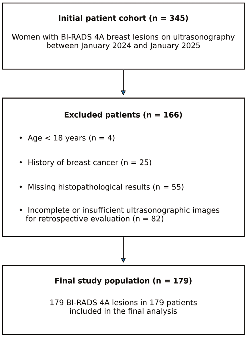
The flowchart about patient selection
BI-RADS: Breast Imaging Reporting and Data System
Ultrasonography Examinations
All examinations were performed using a high-resolution ultrasound system equipped with a linear transducer (central frequency 15 MHz, range 7-18 MHz; Aplio 500; Toshiba Medical Systems, Tokyo, Japan).
Image Evaluation
Ultrasonographic images were independently evaluated by two radiologists with 3 and 8 years’ experience in breast imaging, who were blinded to the patients’ histopathological results, and consensus was achieved in all cases. In cases of disagreement, the opinion of a third radiologist with 25 years of experience was considered.
The evaluated sonographic parameters were defined according to the BI-RADS ultrasound lexicon (2013) () and are summarized in . These parameters included lesion location, size, margin characteristics (circumscribed, indistinct, microlobulated, ill-defined, or spiculated), composition (solid, cystic, or mixed), echogenicity (hypoechoic, isoechoic, or hyperechoic), posterior acoustic features (none, shadowing, or enhancement), and additional findings such as microcalcifications, parenchymal distortion, and ductal ectasia.
Table 1. Distribution of ultrasonographic features of BI-RADS 4A breast lesions according to histopathological subgroup
Feature | Benign (n=149) (%) | High-risk benign (n=18) (%) | Malignant (n=12) (%) | p-value |
|---|---|---|---|---|
Margins Circumscribed | 118 (79%) | 15 (83%) | 4 (33%) | 0.016* |
Irregular | 21 (14%) | 1 (5.5%) | 6 (50%) | |
Microlobulated | 10 (7%) | 2 (11%) | 2 (16%) | |
Microcalcifications | 16 (11%) | 2 (11%) | 5 (42%) | 0.018* |
Parenchymal distortion | 10 (6.7%) | 4 (22%) | 6 (50%) | 0.003* |
Posterior acoustic enhancement | 13 (9%) | 2 (11%) | 0 | 0.748 |
Data are presented as number (percentage). p-values were calculated using the chi-square test or Fisher’s exact test, as appropriate. *Statistically significant (p<0.05). BI-RADS: Breast Imaging Reporting and Data System
Patient age and BI-RADS classification were also recorded. Although mammography reports were available for some patients, they were not consistently present in all cases and were therefore excluded from the comparative analysis. Histopathological diagnoses were categorized into three groups: benign, high-risk benign, and malignant ().
Table 2. Histopathological distribution of BI-RADS 4A breast lesions
Pathology | n (%) |
|---|---|
Benign | 149 (83.3%) |
Fibroadenoma | 81 (54.4%) |
Fibrocystic changes | 51 (34.2%) |
Ductal hyperplasia | 7 (4.7%) |
Mastitis/inflammation | 6 (4.0%) |
Fat necrosis/fibrosis | 4 (2.7%) |
High-risk benign | 18 (10.0%) |
Intraductal papilloma | 11 (61.1%) |
Sclerosing adenosis | 5 (27.8%) |
Phyllodes tumor | 2 (11.1%) |
Malignant | 12 (6.7%) |
Invasive breast carcinoma | 6 (50.0%) |
DCIS | 3 (25.0%) |
Neuroendocrine carcinoma | 1 (8.3%) |
Invasive ductal carcinoma | 1 (8.3%) |
Lobular carcinoma in situ arising from a background of complex fibroadenoma | 1 (8.3%) |
Data are presented as number (percentage) BI-RADS: Breast Imaging Reporting and Data System, DCIS: Ductal carcinoma in situ
Statistical Analysis
Descriptive statistics were expressed as mean ± standard deviation or median (minimum-maximum) for continuous variables and as number and percentage for categorical variables. Normality of data distribution was assessed using the Shapiro-Wilk test.
Continuous variables, including lesion size and age, were compared among groups using the Kruskal-Wallis test. Categorical variables, including margin characteristics, microcalcifications, parenchymal distortion, lesion shape, and posterior acoustic features, were analyzed using the chi-square test or Fisher’s exact test when expected cell frequencies were below 5. Histopathological diagnosis was accepted as the reference standard.
Variables found to be statistically significant in univariate analysis (p<0.05) were further evaluated using multivariate logistic regression analysis to identify independent predictors of malignancy, with histopathological outcome defined as a binary dependent variable (malignant vs. non-malignant, including benign and high-risk benign lesions). Odds ratios (ORs) and 95% confidence intervals (CIs) were calculated. Due to potential multicollinearity between shape and margin characteristics, these variables were not included in the same multivariate model; shape was excluded from the final model. Given the limited number of malignant cases, the multivariate logistic regression analysis was considered exploratory.
Receiver operating characteristic (ROC) curve analysis was performed to evaluate the diagnostic performance of sonographic features identified as significant in the univariate analysis. The area under the curve (AUC), along with sensitivity and specificity, was calculated.
Post-hoc analysis was not performed, as the primary aim of the study was to identify independent predictors of malignancy, which was addressed using multivariate logistic regression.
All statistical analyses were performed using MedCalc Statistical Software version 12.7.7 (MedCalc Software bvba, Ostend, Belgium).
Results
A total of 179 BI-RADS 4A lesions were included in the study. The mean age of the patients was 43±13 years (range: 18-105 years). The mean age was significantly higher in the high-risk benign and malignant groups than in the benign group (p=0.021).
Histopathological diagnoses were categorized as benign (83.3%), high-risk benign (10.0%), and malignant (6.7%), as summarized in . Representative ultrasonographic images from each group are shown in .
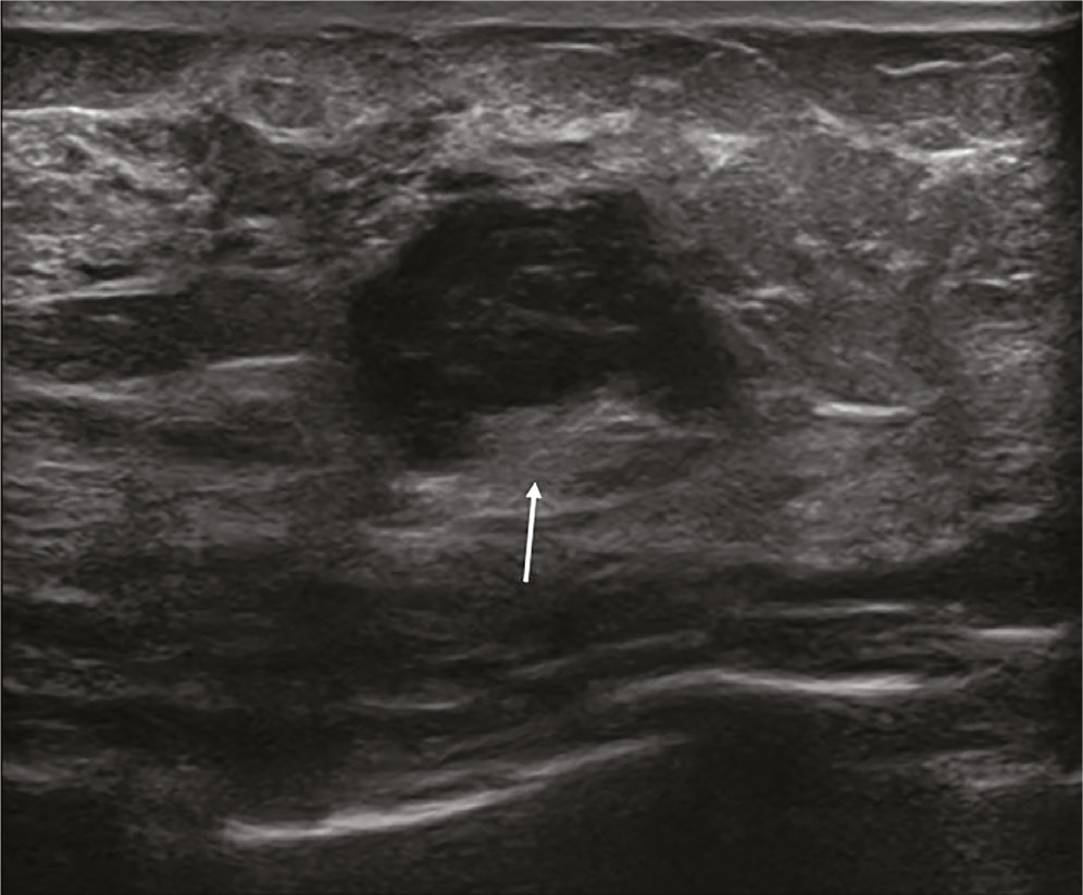
Ultrasound image of a 28-year-old woman demonstrating a hypoechoic solid lesion (indicated by a white arrow) located at the 1 o’clock position of the left breast. The lesion exhibits partially lobulated and partially irregular margins. It was categorized as BI-RADS 4A, and histopathological evaluation confirmed a benign lesion consistent with fibroadenoma
BI-RADS: Breast Imaging Reporting and Data System
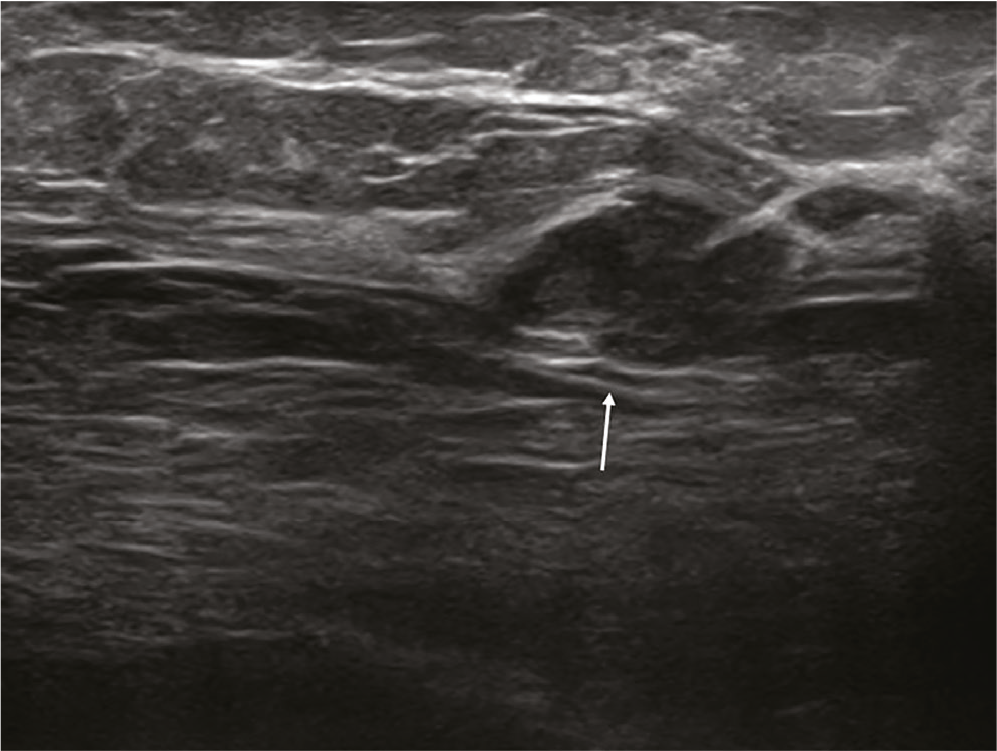
Ultrasound image of a 42-year-old woman demonstrating a lobulated, hypoechoic solid lesion with parallel orientation in the right breast at the 5 o’clock position (indicated by a white arrow). The lesion was categorized as BI-RADS 4A, and histopathological evaluation revealed a high-risk benign lesion consistent with sclerosing adenosis
BI-RADS: Breast Imaging Reporting and Data System
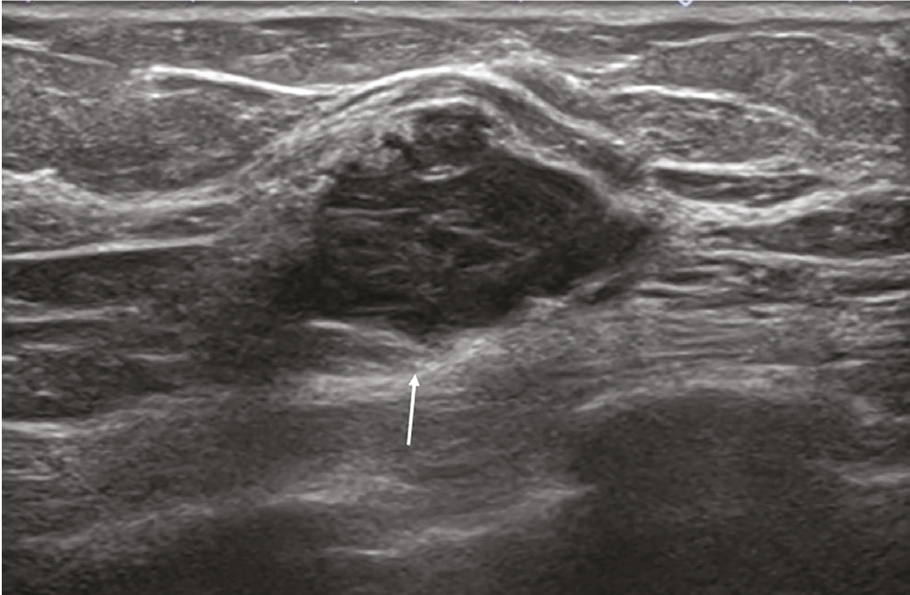
Representative ultrasonographic image of a 55-year-old woman showing a hypoechoic solid lesion with partially indistinct, lobulated, and irregular margins in the left breast at the 2 o’clock position (indicated by a white arrow). The lesion was assessed as BI-RADS 4A on ultrasound, and histopathological examination confirmed a malignant lesion consistent with invasive ductal carcinoma
BI-RADS: Breast Imaging Reporting and Data System
No statistically significant difference was observed in lesion size among the benign, high-risk benign, and malignant groups (Kruskal-Wallis test, χ2=0.48; p=0.787). The mean lesion size was 19.3±11.9 mm in the benign group (range: 3-78 mm), 21.2±17.7 mm in the high-risk benign group (range: 5-63 mm), and 18.8±15.0 mm in the malignant group (range: 4-58 mm).
No statistically significant difference in lesion localization was observed between the groups (chi-square test, p=0.968). Lesions were most frequently located in the upper outer quadrant across all groups, while high-risk benign lesions were relatively predominant in the retroareolar region.
Among the evaluated sonographic features, margin characteristics (p=0.016), microcalcifications (p=0.018), and parenchymal distortion (p=0.003) showed statistically significant differences between the groups (), whereas posterior acoustic enhancement did not differ significantly (p=0.748). Margin irregularity, microcalcifications, and parenchymal distortion were more frequently observed in malignant lesions ().
Lesion shapes differed significantly among the groups (p<0.001). Benign lesions were predominantly oval, whereas irregular shapes were more common in malignant lesions.
Ductal ectasia did not differ significantly between the groups (p=0.61). Similarly, posterior acoustic shadowing showed no significant difference (p=1.00).
Composition (p=0.928) and echogenicity (p=0.546) also did not differ significantly between the groups.
Variables found to be significant in univariate analysis were further evaluated using multivariate logistic regression. In multivariate analysis, the presence of microcalcifications was identified as an independent predictor of malignancy (OR: 6.51, 95% CI: 1.66-25.52, p=0.007). Although parenchymal distortion (OR: 4.18, 95% CI: 0.57-30.63, p=0.16) and margin characteristics (OR: 2.28, 95% CI: 0.55-9.40, p=0.25) showed increased ORs, neither remained statistically significant in the multivariate model.
Receiver operating characteristic curve analysis was performed to evaluate the diagnostic performance of sonographic features. Microcalcifications demonstrated the highest diagnostic performance (AUC=0.66, 95% CI: 0.50-0.82), followed by lesion shape (AUC=0.64, 95% CI: 0.48-0.80), parenchymal distortion (AUC=0.60, 95% CI: 0.43-0.77), and margin characteristics (AUC=0.56, 95% CI: 0.40-0.72). The overall malignancy rate of BI-RADS 4A lesions in this cohort was 6.7%.
Discussion
This study provides a focused evaluation of sonographic predictors of malignancy in BI-RADS 4A breast lesions, a subgroup characterized by considerable diagnostic uncertainty. Several sonographic features, including irregular margins, irregular shape, parenchymal distortion, and microcalcifications, were significantly associated with malignancy in univariate analysis. However, in multivariable analysis, microcalcifications remained associated with malignancy; this finding should be interpreted cautiously given the limited number of malignant events. These results highlight the limitations of relying solely on conventional sonographic descriptors and emphasize the necessity of more refined risk-stratification strategies. From a clinical perspective, these findings may contribute to risk stratification within the BI-RADS 4A category.
The reported malignancy rate for BI-RADS 4A lesions ranges from 2% to 10% in the literature (,). The malignancy rate observed in our study (6.7%) falls within this range, supporting the representativeness of our cohort. This finding reinforces the notion that although the risk of malignancy is relatively low, BI-RADS 4A lesions remain clinically significant and require careful evaluation.
Advanced age has consistently been identified as a predictor of malignancy in BI-RADS 4A lesions (). In our study, the higher mean age observed in the malignant and high-risk benign groups further supports this association. From a clinical perspective, these results suggest that patient age may be a complementary parameter for risk stratification.
The association between lesion size and malignancy has been reported inconsistently in previous studies (,). However, lesion size alone is not considered a reliable discriminative feature (). Consistent with these findings, no significant association between lesion size and malignancy was observed, supporting the limited discriminative value of this parameter. Therefore, lesion size should not be considered an independent determinant in clinical decision-making. Our findings further underscore the complexity of risk stratification in BI-RADS 4A lesions.
The ultrasonographic appearance of breast lesions can be highly variable, and both benign and malignant features may coexist within the same lesion (,). This heterogeneity contributes to diagnostic uncertainty, particularly in BI-RADS 4A lesions, where overlapping imaging findings may lead to misclassification. In addition, interobserver variability in interpreting BI-RADS descriptors further complicates lesion categorization (). These factors collectively highlight the inherent limitations of conventional sonographic assessment and underscore the need for more standardized, reproducible evaluation approaches.
Irregular shape is a recognized feature associated with malignancy in breast lesions (,). However, it was not included in the multivariate model due to its strong association with margin characteristics, which may have introduced collinearity and affected model stability. Therefore, its independent predictive contribution could not be determined in this study.
Margin characteristics are well-established features associated with malignancy in breast lesions (). Margin irregularity was more frequently observed in malignant lesions, supporting its role in the initial assessment of malignancy. Although margin characteristics were significantly associated with malignancy in univariate analysis, they did not remain independently significant in multivariate analysis. These results suggest that margin characteristics alone may be insufficient for accurate risk stratification.
Echogenicity has limited discriminative value in distinguishing benign from malignant breast lesions, as substantial overlap exists between groups (). In the present study, no significant difference in echogenicity was observed, which further supports its limited discriminative value in malignancy prediction.
Cystic morphology is generally associated with benign breast lesions, whereas solid and mixed patterns have limited discriminative value (,). In our study, cystic morphology was observed exclusively in benign lesions. In contrast, solid and mixed morphologies were similarly distributed across groups, suggesting a limited role for lesion composition in malignancy differentiation.
Posterior acoustic shadowing is commonly considered a feature suggestive of malignancy; however, it may also be observed in benign lesions, particularly in the presence of fibrosis or calcifications (). In our study, posterior acoustic features did not show a significant association with malignancy, a finding that may be partly explained by the limited number of malignant cases.
Microcalcifications are well-recognized as a feature associated with malignancy in breast lesions (,,). In our study, microcalcifications were the only parameters that retained independent predictive value in multivariate analysis, highlighting their importance among the evaluated sonographic features. This finding supports the role of microcalcifications as a more reliable feature in BI-RADS 4A lesions compared to other conventional descriptors.
Although microcalcifications were the only feature that remained independently associated with malignancy in the multivariate analysis, they demonstrated moderate diagnostic performance in ROC analysis (AUC=0.66). The lower discriminatory performance of the other sonographic features, with AUC values ranging from 0.56 to 0.64, suggests that microcalcifications, despite their moderate diagnostic performance, may contribute relatively more to risk stratification. From a clinical perspective, the independent association between microcalcifications and malignancy suggests that their presence may serve as an additional indicator of increased malignancy risk in BI-RADS 4A lesions, particularly when accompanied by other suspicious sonographic findings. Nevertheless, given their moderate diagnostic performance, microcalcifications should not be considered a standalone determinant of biopsy decisions but rather an additional sonographic feature that may support risk assessment within the BI-RADS 4A category.
Recent studies have explored advanced approaches to improve diagnostic accuracy in BI-RADS 4 lesions, including multimodal ultrasound, elastography, contrast-enhanced ultrasound, deep learning, and nomogram-based models (,,,). While these approaches aim to enhance overall diagnostic performance, they also underscore the limitations of relying solely on individual conventional sonographic features. In this context, our findings indicate that certain features, particularly microcalcifications, may retain a more robust predictive role than other descriptors.
Orientation is generally considered a supportive feature in lesion assessment, with benign lesions more commonly aligned parallel to the skin (). In our study, however, orientation alone was not a distinctive parameter, suggesting that its contribution to malignancy prediction may be limited when evaluated in isolation.
Parenchymal distortion has been associated with malignancy due to its relation to stromal retraction and infiltrative growth patterns (). In our study, distortion was more frequently observed in malignant lesions; however, it did not retain statistical significance in multivariate analysis, suggesting that other accompanying features may influence its predictive value.
Previous studies have reported that the most common benign diagnoses in the BI-RADS 4A category are fibroadenoma and fibrocystic changes (,). Consistent with these findings, the majority of histopathological diagnoses in our study were fibroadenomas and fibrocystic changes.
Intraductal papilloma was the most common lesion within the high-risk benign group, followed by sclerosing adenosis. These lesions may exhibit imaging features overlapping with malignancy, including irregular margins, microcalcifications, and parenchymal distortion (,), potentially contributing to false-positive classification in BI-RADS 4A lesions. Among malignant cases, invasive ductal carcinoma was the most common diagnosis, reflecting its known predominance among breast cancers ().
Study Limitations
Our study has several limitations. The retrospective single-center design limits the generalizability of the results. In addition, only ultrasonographic findings were evaluated; vascularity and elastographic features of the lesions were not assessed. Breast parenchymal characteristics, including tissue density, were also not evaluated, which may have affected lesion assessment accuracy and contributed to potential misclassification. As the US is an operator-dependent technique with substantial interobserver variability, differences in experience and the learning curve may lead to classification inconsistencies (). Furthermore, evaluating static images may have led to overlooking dynamic information that real-time US could provide.
Although ultrasonographic images were independently evaluated by two radiologists, only the final consensus assessments were used for analysis. Consequently, interobserver agreement could not be formally assessed using Cohen’s kappa or the intraclass correlation coefficient, which constitutes a limitation of the present study.
Another limitation is that benign cases diagnosed by core biopsy were not confirmed by surgical excision or long-term follow-up, potentially resulting in false-negative outcomes. The literature reports a false-negative rate of approximately 3.7% for 14-gauge US-guided core biopsies (). Because certain sonographic variables were strongly associated, not all features could be included simultaneously in the multivariate model, which may have limited the ability to fully assess the independent predictive contributions of these variables.
Furthermore, the limited number of malignant cases may have affected the stability of the multivariate logistic regression model and increased the risk of overfitting. Therefore, the identified independent predictor should be interpreted with caution and validated in larger, independent cohorts.
Despite these limitations, this study has several strengths. First, all sonographic evaluations were performed independently by radiologists blinded to histopathological results using standardized BI-RADS 2013 ultrasound descriptors, followed by consensus assessment, thereby minimizing potential interpretation bias. Second, histopathological confirmation was available for all included lesions, and no patients were lost to follow-up, ensuring complete diagnostic verification. Third, the detailed categorization of lesions into benign, high-risk benign, and malignant groups reflects real-world clinical practice and provides a clinically relevant framework. Fourth, multivariate analysis was performed after a careful assessment of multicollinearity, allowing the robust identification of independent predictors of malignancy. The observed malignancy rate (6.7%) falls within the reported range for BI-RADS 4A lesions (2-10%), supporting the representativeness of the study sample.
Conclusion
Microcalcifications emerged as the only independent ultrasonographic predictor of malignancy in BI-RADS 4A breast lesions. Although irregular shape, margin characteristics, and parenchymal distortion were significantly associated with malignancy in univariate analysis, only microcalcifications retained independent significance in multivariate analysis. These findings suggest that conventional sonographic descriptors may have limited standalone discriminative value. Emphasizing more reliable imaging features, particularly microcalcifications, may contribute to malignancy risk stratification in BI-RADS 4A lesions.
However, these findings require validation in larger, multicenter prospective studies to confirm their generalizability and clinical applicability.
Ethics
Ethics Committee Approval: The study was approved by the Ethics Committee of Umraniye Training and Research Hospital (approval number: 27.02.2025/19, date: 27.02.2025) and was conducted in accordance with the ethical principles outlined in the Declaration of Helsinki.
Informed Consent: Due to the study’s retrospective design, the ethics committee waived the requirement for informed consent.
Footnotes
Authorship Contributions
Concept: G.G., F.K., Design: G.G., S.N.A., Data Collection or Processing: G.G., S.N.A., Analysis or Interpretation: G.G., F.K., Literature Search: G.G., Writing: G.G., F.K.
Conflict of Interest
The authors declared that there were no conflicts of interest.
Financial Disclosure
The authors declared that this study received no financial support.
References
Siegel RL, Giaquinto AN, Jemal A. Cancer statistics, 2024. CA Cancer J Clin. 2024;74:12-49.
Duggan SN, Azharuddin M, Hernández R, et al. Supplemental imaging modalities for breast cancer screening in women with dense breasts: a systematic review with economic considerations. Breast. 2026;85:104668.
Gilbert FJ, Payne NR, Allajbeu I, et al. Comparison of supplemental breast cancer imaging techniques-interim results from the BRAID randomised controlled trial. Lancet. 2025;405:1935-44.
American College of Radiology. ACR BI-RADS® Atlas—Breast Imaging Reporting and Data System. Reston (VA): American College of Radiology; 2025.
Pradeep Kumar D, Chakiath AX, Sugunan A, et al. Rethinking breast imaging reporting and data system (BI-RADS) 4: clinical insights from mammographic subclassification. Cureus. 2025;17:e94379.
Castro-Palomares JE, Sanchez-Rodriguez IG, Reyes-Dircio A, et al. Correlation between BI-RADS 4 subcategories and histopathological outcomes in Mexican women with breast lesions: a retrospective study of lesion laterality and cancer incidence. Curr Med Imaging. 2025;21:e15734056407923.
Zhu Y, Jiang L, Chen YN, et al. Diagnostic value of real-time ultrasound elastography and contrast-enhanced ultrasound in BI-RADS 4A breast lesions. Gland Surg. 2025;14:488-97.
Li C, Wu M, Haiqing H. Application of multimodal ultrasound examination in BI-RADS category 4 breast lesions. Front Med (Lausanne). 2025;12:1596100
Zhong Y, Chen YT, Qiu YD, et al. Sonographic glandular tissue component: a potential imaging marker for upgrading BI-RADS 4A breast masses. Acad Radiol. 2025;32:3883-91.
Wang J, Ma H, Wang S, et al. Optimizing S-detect classification accuracy for BI-RADS 4 breast nodules using multimodal ultrasound parameters. Quant Imaging Med Surg. 2026;16:159.
American College of Radiology. ACR BI-RADS® Atlas: Breast Imaging Reporting and Data System. 5th ed. Reston(VA):American College of Radiology; 2013.
Hu Y, Yang Y, Gu R, et al. Does patient age affect the PPV3 of ACR BI-RADS ultrasound categories 4 and 5? Eur Radiol. 2018;28:2492-8.
Lazarus E, Mainiero MB, Schepps B, et al. BI-RADS lexicon for US and mammography: interobserver variability and positive predictive value. Radiology. 2006;239:385-91.
Ghunaim HA. Effect of patient age and breast parenchymal density on BI-RADS 4 subcategorization. Pak J Med Sci. 2024;40:2356-62.
Elverici E, Barca AN, Aktas H, et al. Nonpalpable BI-RADS 4 breast lesions: sonographic findings and pathology correlation. Diagn Interv Radiol. 2015;21:189-94.
Bouzghar G, Levenback BJ, Sultan LR, et al. Bayesian probability of malignancy with BI-RADS sonographic features. J Ultrasound Med. 2014;33:641-8.
Wiratkapun C, Bunyapaiboonsri W, Wibulpolprasert B, et al. Biopsy rate and positive predictive value for breast cancer in BI-RADS category 4 lesions. J Med Assoc Thai. 2010;93:830-7.
Stavros AT, Thickman D, Rapp CL, et al. Solid breast nodules: use of sonography to distinguish between benign and malignant lesions. Radiology. 1995;196:123-34.
Mohapatra SK, Mishra A, Sahoo TK, et al. Positive predictive values of BI-RADS 4 lesions and its mammographic features. Indian J Surg Oncol. 2021;12:182-9.
Tan YY, Wee SB, Tan MP, et al. Positive predictive value of BI-RADS categorization in an Asian population. Asian J Surg. 2004;27:186-91.
Chaitanya INVL, Prabhala S, Srirambhatla A, et al. Comparison of histopathologic findings with BI-RADS score in tru-cut biopsies of breast lesions. Int J Pathol Res Pract. 2020;9:35-41.
Hong AS, Rosen EL, Soo MS, et al. BI-RADS for sonography: positive and negative predictive values. AJR Am J Roentgenol. 2005;184:1260-5.
Costantini M, Belli P, Lombardi R, et al. Characterization of solid breast masses using the sonographic BI-RADS lexicon. J Ultrasound Med. 2006;25:649-61.
Brunetti N, Campi C, Piana M, et al. A radiomic and clinical data-based risk model for malignancy prediction of breast BI-RADS 4A microcalcifications. Clin Breast Cancer. 2025;25:e461-9.
Yang Y, Liao T, Lin X, et al. Reducing unnecessary biopsies of BI-RADS 4 lesions using deep learning in mammography. Front Oncol. 2025;15:1543553.
Jiang X, Chen C, Yao J, et al. Nomogram for diagnosis of BI-RADS 4 breast nodules based on 3D ultrasound. BMC Med Imaging. 2025;25:48.
Shen L, Jiang T, Tang P, et al. Quantitative malignant risk prediction of amorphous calcifications. Quant Imaging Med Surg. 2022;12:2672-83.
Schoonjans JM, Brem RF. Fourteen-gauge ultrasound-guided core needle biopsy of breast masses. J Ultrasound Med. 2001;20:967-72.
Lee YJ, Choi SY, Kim KS, et al. Observer variability using BI-RADS ultrasound (5th edition). Iran J Radiol. 2016;13:e28281.
Crystal P, Koretz M, Shcharynsky S, et al. Accuracy of ultrasound-guided core needle biopsy of breast lesions. J Clin Ultrasound. 2005;33:47-52.