
Abstract
Aim
Transcutaneous electrical stimulation (TES) is widely used in clinical settings and can produce both direct and indirect effects on the sinoatrial node (SAN). This study aimed to investigate whether TES affects SAN function directly or indirectly via the autonomic nervous system.
Methods
The study involved 24 young, healthy adult volunteers and was conducted in two phases. Experiment 1 examined the direct effects of TES on the SAN, and Experiment 2 examined its indirect effects. In Experiment 1, which used a single-pulse electrical current, participants underwent real and sham TES sessions in random order. The P-QRS response and heart rate variability (HRV) in both time and frequency domains were assessed. In Experiment 2, in which a pulse-train electric current was used, conventional and acupuncture-like transcutaneous electrical nerve stimulation were applied in random order, and HRV was analyzed similarly. In both experiments, the electrical current was applied to the volar aspect of the forearm.
Results
Peristimulus time histograms and waveform averages revealed no heartbeat associated with the electrical pulse. Additionally, time and frequency domain analyses indicated that pulse-train TES had no effect on HRV.
Conclusion
This study is the first to examine the direct effects of TES on SAN action potential and HRV. Results show that low-frequency TES has no direct or indirect impact on heart rhythm or SAN function in healthy individuals and confirm its safety for diagnostic and therapeutic use.
Keywords: Electrical stimulation, transcutaneous electric nerve stimulation, heart rate, tachycardia, bradycardia
Introduction
Transcutaneous electrical stimulation (TES) has been a staple in clinical practice for over a century, used to diagnose and treat various medical conditions. Therapeutic TES, delivered in pulse-train form, is commonly employed in physical therapy, rehabilitation, and professional sports; modalities include neuromuscular electrical stimulation, functional electrical stimulation, and transcutaneous electrical nerve stimulation (TENS) (,). Additionally, diagnostic TES, typically in single-pulse form, is frequently used in electromyographic reflex studies to diagnose neuromuscular disorders (). Although the benefits of TES are well known, its potential side effects on the heart have yet to be convincingly clarified.
The heart’s intrinsic pacemaker, the sinoatrial node (SAN), rhythmically generates action potentials propagating through the myocardium, causing the heart to contract. The function of the SAN is modified by the sympathetic and parasympathetic nerve fibers. Electrical current is suggested to indirectly change SAN rhythm through these nerve fibers (,). These effects likely dissipate after cessation of electrical stimulation. However, previous observations were conducted several minutes after electrical stimulation rather than during it, resulting in conflicting findings (-). In some studies, it has been reported that heart rate variability (HRV) decreases (,); in others, it increases or decreases depending on the frequency of the electrical stimulus (), while in others, it does not change ().
We hypothesized that electrical current may directly and immediately affect SAN excitability. Owing to the high conduction speed of electrical current, such effects, if present, would be expected to occur within a very short latency window and therefore be temporally locked to the stimulus (i.e., time-locked, synchronous responses). In other words, if TES directly influences action potential generation in the SAN, an electrical stimulus-related P-QRS complex should appear immediately after the stimulus. Such stimulus-locked cardiac responses can be detected using peristimulus time histograms (PSTHs) and waveform-averaging analyses. Unlike previous studies that evaluated heart rate or HRV before and after stimulation—thus primarily reflecting delayed autonomic modulation—the present study specifically tests the immediate, stimulus-synchronous (direct) effect of TES on SAN activity in humans while also examining potential indirect autonomic influences. These findings, by clarifying the cardiac safety profile of TES, may contribute to the safer clinical application of electrotherapy in vulnerable patient populations.
Materials and Methods
Compliance with Ethical Standards
All volunteers provided written informed consent to the experimental procedures, which adhered to the principles outlined in the Declaration of Helsinki and were approved by the Koç University Local Ethics Committee (protocol no: 2017.124.IRB2.038, date: 05.09.2017). Ethical approval was obtained from Koç University because one of the authors, Prof. Dr. Kemal Sitki Turker, was a faculty member at the Koç University at the time the study protocol was approved. Ethical approval was obtained from Koç University because one of the authors, Prof. Dr. Kemal Sitki Turker, was a faculty member at the Koç University at the time the study protocol was approved. The study protocol was registered with ClinicalTrials.gov (NCT05966922, July 13, 2023). This research comprised two phases: Experiment 1 analyzed the impact of a single electrical pulse on HRV, while Experiment 2 explored the effects of TES delivered as pulse trains on HRV.
Study Design and Participants
Healthy volunteers aged 20-45 were recruited for the study; exclusion criteria included pregnancy, alcohol or drug abuse, and use of any medication affecting the cardiovascular system. Twelve male volunteers took part in Experiment 1, and twelve volunteers (one female and eleven males) participated in Experiment 2. The mean ages of participants in Experiments 1 and 2 were 26.3±2.9 and 25.5±6.5 years, respectively. These two experiments were conducted using separate groups to manage the lengthy and complex experimental procedures effectively.
Experimental Procedures
The experimental design, including electrode placement on the forearm and hypothesized neural pathways, is illustrated in . Participants rested for 3 minutes in a quiet, temperature-controlled room maintained at 23 °C before each experiment. They were asked to abstain from strenuous exercise and caffeine consumption for at least 2 hours. Additionally, participants were advised to refrain from activities such as talking, coughing, and deep breathing during measurements to minimize potential influences on the results.
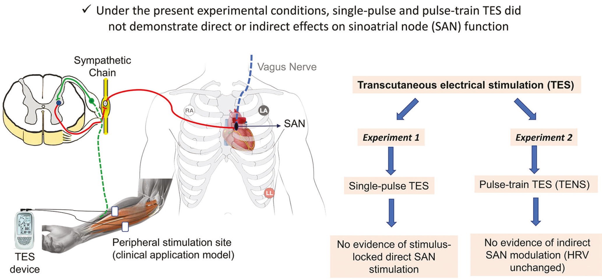
Experimental design and investigated neural pathways. TES was applied to the forearm to investigate potential direct and indirect effects on SAN function. The diagram illustrates the hypothesized neural pathways -the sympathetic chain (red) and the vagus nerve (blue)- and the two-phase experimental design. Experiment 1 tested for direct electrical effects using single-pulse stimulation, while Experiment 2 assessed indirect autonomic effects using pulse-train stimulation
During electrical stimulation, participants were seated with their elbows flexed at 90 degrees and their hands supinated. Self-adhesive electrodes (4 x 4 cm) were positioned on the flexor aspect of the right forearm to deliver the electrical current (). Heart rate variability changes during rest and stimulation were monitored with a three-lead electrocardiogram (ECG).
Experiment 1 (Testing a Single Electrical Pulse on Heart Rate Variability)
Participants underwent both TES and Sham TES sessions in random order. Each session lasted 600 seconds, with a 5-minute rest interval between sessions. Sham TES was performed to observe random variations in the occurrence of the P-QRS complex over time.
Throughout the experiment, the software (Spike2) used the peak of the R wave within the P-QRS complex observed in the ECG channel as a trigger. This triggering allowed the delivery of an electrical stimulus within a 20-250 ms interval before the next P-wave. Trial recordings substantiated that the electrical stimulation consistently occurred 20-250 ms before the onset of the P-wave.
A single monophasic square electrical pulse (i.e., unidirectional pulsed current, also termed interrupted direct current/galvanic) was administered at random intervals of 1 to 4 seconds using a constant-current stimulator (model DS7A, Digitimer Ltd, Hertfordshire, UK) with a pulse width of 200 microseconds. Participants received the highest current intensity they could tolerate, with a mean intensity of 48.1±13.3 milliamperes (mA), ranging from 30 to 70 mA. During the sham stimulation session, the current intensity was set to zero mA.
Experiment 2 (Testing Transcutaneous Electrical Nerve Stimulation on Heart Rate Variability)
In this experiment, conventional and acupuncture-like TENS, which are commonly used in clinical practice, were employed as pulse-train TENS modalities. Initially, a resting ECG was recorded for 800 seconds. Subsequently, one session of conventional TENS and one session of acupuncture-like TENS were applied in random order. Each TENS session lasted 800 seconds, with a 5-minute rest interval between sessions.
The conventional mode is characterized by a high-frequency (HF) current (100 Hz, 200 µs pulse width, 4-second on period, and 10-second off period). The stimulus intensity was adjusted to a level at which the subject experienced minimal paresthesia without muscle contractions. If the tingling sensation faded, the current intensity was increased until the sensation was perceived again. The acupuncture-like mode is characterized by a low frequency (LF) current (10 Hz, 200 µs pulse width, 4 seconds on period, 10 seconds off period). The stimulus intensity was adjusted to a level that allowed for comfortable contractions of the wrist flexors. TENS was administered using the CefarRehab® X2 device (DJO France SAS, Mouguerre, France).
Three-lead ECG Recording
The participant’s ECG data were continuously recorded using non-invasive sensors (self-adhesive pre-gelled Ag/AgCl electrodes, Redline® Istanbul, Turkiye) with standard three-lead ECG chest placement. Specifically, the active electrode was placed at the right second intercostal space and the reference electrode at the left second intercostal space, both along the midclavicular line. Another active electrode was situated on the anterior chest wall at the left lower rib cage. Before electrode placement, the skin was shaved, lightly abraded, cleaned with alcohol, and moistened with ECG gel. Skin resistance was measured to ensure an optimal connection (less than 10 kiloohms) ().
ECG signals were recorded using a data acquisition system (CED 1902 Quad System MKIII amplifier and CED 3601 Power 1401 MKII digital-to-analog converter, Cambridge Electronic Designs, Cambridge, UK). The ECG potentials were amplified 100 times and recorded at a sampling frequency of 2 kHz.
Outcome Variables and Recording Analysis
The primary outcome variables differed between the two experiments because they addressed distinct physiological mechanisms and time scales of potential TES effects on the SAN. In Experiment 1, the main outcome was the electrical stimulus-related QRS response, assessed using PSTHs and waveform-averaging analyses to determine whether TES could evoke an immediate, stimulus-locked cardiac response indicative of a direct effect on SAN excitability. Beat-to-beat RR interval changes (i.e., HRV) were analyzed to explore short-term variability accompanying single-pulse stimulation.
In contrast, Experiment 2 was designed to evaluate the possible indirect autonomic influence of pulse-train TES on SAN function over time; therefore, the primary outcome was HRV derived from RR interval fluctuations in the time and frequency domains. Thus, while both experiments involved ECG-derived measures, Experiment 1 focused on event-related, stimulus-synchronous QRS responses, whereas Experiment 2 examined slower, autonomically mediated modulation of cardiac rhythm, reflected in HRV.
The Electrical Stimulus-Related QRS Response
If TES affects cardiac rhythm, it would be expected to advance or delay the timing of the QRS response. The possible direct effect of an electrical stimulus on the timing of the QRS response was investigated in the time domain using the PSTH and waveform averaging (). If a typical P-QRS complex, similar to the one just before stimulation, was evident in the waveform-averaged graphs after electrical stimulation, or if a very narrowly distributed bell curve appeared in the PSTH graphs compared with sham, the observation was interpreted as an electrical current-related P-QRS complex.
Heart Rate Variability
The time-domain analysis assessed beat-to-beat interval variability (i.e., R-R interval differences). This analysis included the standard deviation (SD) of the R-R intervals (SDRR), the coefficient of variation of the R-R intervals (CVRR), and the square root of the mean of the squared differences between adjacent R-R intervals (RMSSD). Prolongation of the R-R interval or an increase in R-R interval variability (SDRR, CVRR, and RMSSD) was interpreted as an increase in parasympathetic tone, reflecting greater parasympathetic (vagal) modulation of cardiac rhythm, which in healthy individuals is generally associated with more adaptive autonomic regulation ().
Frequency-domain HRV was analyzed in accordance with the guidelines of the Task Force of the European Society of Cardiology and the North American Society of Pacing and Electrophysiology. The fast fourier transform was used to calculate the LF (0.04-0.15 Hz) and HF (0.15-0.4 Hz) components. Low-frequency and HF components were considered to reflect sympathetic and parasympathetic activity, respectively. The sympathovagal balance was assessed by the LF/HF ratio, where ratios greater than 1 indicated sympathetic dominance (sympatheticotonia), ratios less than 1 indicated parasympathetic dominance (vagotonia), and a ratio of 1 indicated sympathovagal balance (). ECG data were analyzed using the Spike2 7.20 software package (CED, UK).
Statistical Analysis
An a priori power analysis was conducted using G*Power (version 3.1.9.7) for a paired-samples t-test. The sample size was calculated based on the study by Ngai and Jones (), which reported an effect size of 1.25 for the LF variable. This effect size was considered appropriate because the present study was designed to detect clinically meaningful alterations in HRV rather than subtle physiological fluctuations. Assuming a power of 95% and an alpha error of 5%, the required sample size was 11 subjects per experiment.
The normality of the data distribution was assessed using the Shapiro-Wilk test. Data were summarized as arithmetic means and SDs. Paired t-tests or repeated-measures analysis of variance (ANOVA) were used to compare the mean values across experimental sessions. Post-hoc pairwise comparisons with Bonferroni correction were performed only when the overall repeated-measures ANOVA yielded a statistically significant effect. Categorical dependent variables were compared using Cochran’s Q test for related samples. Statistical significance was set at p<0.05. Data management and analysis were conducted using PASW Statistics for Windows, version 18.0 (SPSS Inc., Armonk, NY).
Results
Data analysis from Experiment 1, using PSTHs and waveform averages, indicated no heartbeat related to the electrical pulse. Waveform-averaging analysis revealed that a typical P-QRS complex did not occur in any of the participants after electrical stimulation. No difference was detected in PSTH histogram patterns between electrical stimulation and sham stimulation (). Furthermore, there were no statistically significant differences between electrical and sham stimulation for HRV variables in both the time and frequency domains ().
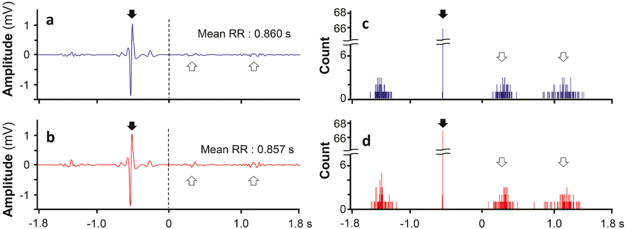
Waveform average plots (a, b) and peristimulus time histograms (PSTH; c, d) from a representative subject. The upper panels (a, c) correspond to sham stimulation, whereas the lower panels (b, d) correspond to real stimulation. Vertical dashed lines indicate the timing of electrical pulse delivery. P-QRS complexes immediately preceding stimulation are marked with solid arrows, whereas those occurring after stimulation are indicated with open arrows. No stimulus-locked P-QRS complex was observed following electrical stimulation, and the PSTH distributions were comparable between the sham and real stimulation conditions
Table 1. Heart rate variability in the time and frequency domain in Experiment 1
- | TES | Sham TES | p-value | |
|---|---|---|---|---|
Average RR (ms) | 762.6±70.4 | 749.8±70.6 | 0.095 | |
SDRR (ms) | 47.2±12.0 | 50.6±12.3 | 0.081 | |
CVRR (%) | 6.3±1.5 | 6.7±1.7 | 0.104 | |
RMSSD (ms) | 28.5±9.8 | 28.2±8.3 | 0.857 | |
Low frequency (nu) | 75.5±9.2 | 78.1±7.8 | 0.353 | |
High frequency (nu) | 25.0±8.5 | 22.0±7.5 | 0.225 | |
Low/High frequency ratio | 3.5±1.6 | 4.0±1.6 | 0.249 | |
Low/High frequency (N) | >1 | 12 | 12 | NS |
<1 | 0 | 0 | ||
Data are presented as mean ± standard deviation. p-values were calculated using a paired-samples t-test. Statistical significance was set at p<0.05 NS: Non-significant, nu: Normalized units, N: Number of subjects, ms: Millisecond, SDRR: Standard deviation of the R-R intervals, CVRR: Coefficient of variation of the R-R intervals, RMSSD: Square root of the mean of the sum of squares of differences between adjacent R-R intervals, TES: Transcutaneous electrical stimulation
Analysis of Experiment 2 data revealed no statistically significant differences in time-domain and frequency-domain HRV variables among the resting state, conventional TENS, and acupuncture-like TENS (). Although LF, HF, and the LF/HF ratio showed borderline p-values in the repeated-measures ANOVA, no post-hoc pairwise comparisons were conducted because the overall ANOVA did not reach statistical significance.
Table 2. Effects of TENS on the heart rate variability in the time and frequency domain
- | Rest | Acupuncture like TENS | Conventional TENS | p-value | Partial eta squared | |
|---|---|---|---|---|---|---|
Average RR (ms) | 740.3±67.5 | 741.0±63.0 | 755.3±66.8 | 0.161* | 0.161 | |
SDRR (ms) | 47.8±17.3 | 46.9±7.5 | 46.7±11.8 | 0.873* | 0.006 | |
CVRR (%) | 6.4±2.1 | 6.3±0.8 | 6.2±1.5 | 0.845* | 0.015 | |
RMSSD (ms) | 27.4±11.4 | 27.8±8.1 | 29.3±10.5 | 0.444* | 0.061 | |
Low frequency (nu) | 64.7±12.4 | 72.3±9.8 | 72.9±9.2 | 0.054* | 0.253 | |
High frequency (nu) | 34.9±11.1 | 27.7±9.5 | 27.0±8.5 | 0.057* | 0.263 | |
Low/High frequency ratio | 2.2±1.1 | 3.1±1.7 | 3.1±1.4 | 0.057* | 0.249 | |
Low/High frequency (N) | >1 | 11 | 12 | 12 | 0.368** | - |
<1 | 1 | 0 | 0 | - | ||
*Repeated measures of variance analysis, **Related-samples Cochran’s Q test TENS: Transcutaneous electrical nerve stimulation, SDRR: Standard deviation of the R-R intervals, CVRR: Coefficient of variation of the R-R intervals, RMSSD: Square root of the mean of the sum of squares of differences between adjacent R-R intervals
Discussion
This study, which included young, healthy adult volunteers, yielded two original findings: PSTH and waveform-average analyses of ECG data recorded during the TES session showed that the electrical current had no direct effect on SAN function. Secondly, HRV analysis of ECG data recorded during the TES session showed that the electrical current had no indirect effect on SAN function via the autonomic nervous system (ANS).
Importantly, this methodological distinction has critical implications for interpreting the existing literature. Studies relying on pre-post HRV comparisons may capture delayed or non-specific autonomic adjustments rather than the immediate physiological response to stimulation (-). By contrast, the present study isolates both stimulus-synchronous electrophysiological responses and real-time autonomic modulation during TES. Therefore, the absence of both a stimulus-locked P-QRS response and a concurrent HRV change provides converging evidence that TES, under the present conditions, does not exert a functionally meaningful influence on SAN activity. Importantly, this should be interpreted as evidence of absence within the temporal resolution and sensitivity of the applied methods, rather than a definitive exclusion of all possible effects under different stimulation conditions.
To assess the effect of TES on sinus node function via the ANS, we did not apply stimulation to sympathetic ganglia, such as the stellate ganglion, in this study. Instead, TES was delivered to the forearm, consistent with its common peripheral application in clinical settings. This approach was chosen to reflect typical TES usage and to avoid direct stimulation of cardiac-related autonomic structures.
To directly stimulate the SAN, an electrical pulse must reach it before the onset of the atrial action potential’s absolute refractory period. In Experiment 1, an electrical pulse locked to the R-wave was delivered to ensure this. Specifically, the R-wave was used as the trigger, and a single electrical pulse was delivered within an interval of 20-250 ms before the subsequent P-wave. ECG recordings obtained during TES were analyzed using PSTH and waveform-averaging techniques to detect the stimulus-related P-QRS response; this response was then compared with responses obtained during sham stimulation.
The SAN function is modulated by both sympathetic and parasympathetic nerve fibers (). Experiment 2 tested the indirect effect of TES on the SAN via the ANS. HRV, a non-invasive measure of ANS activity, was used as an objective index for assessment (). While previous studies typically analyzed HRV before and after TES, this study analyzed HRV from ECG recordings obtained during both rest and TENS sessions, which distinguishes it from prior research. This methodological distinction is critical, as post-stimulation HRV measurements may reflect residual autonomic responses, relaxation effects, or postural changes rather than the direct neuromodulatory effects of TES.
During Experiment 1, no stimulus-locked, pulse-related P-QRS response was observed, indicating that the electrical current did not directly excite the SAN. While previous TES studies have primarily evaluated delayed changes in heart rate or HRV reflecting autonomic modulation, the present study specifically examined the immediate, stimulus-synchronous electrophysiological response of the SAN. To our knowledge, this time-locked approach to testing a direct effect of TES on the SAN has not previously been reported in humans.
The alpha motor neuron regulates the functions of skeletal muscle. In electrophysiological studies, the alpha motor neuron or its axon is stimulated with TES, and a skeletal muscle response associated with an electric pulse is observed in electromyography (). Similarly, if the SAN can be stimulated with TES, a P-QRS complex associated with an electrical pulse is expected to occur on the ECG. PSTH and waveform averaging are commonly used methods to show stimulus-related responses (). In Experiment 1, ECG recordings taken during TES did not reveal a P-QRS response associated with an electrical pulse. illustrates that the histogram and P-QRS patterns generated using the PSTH and waveform-average graphs of the ECG recordings taken during real and sham stimulations are remarkably similar. These analyses also revealed that a typical P-QRS wave was not observed after TES. These findings indicate that TES does not have a direct effect on the SAN.
It has been observed that TES activates the ANS, as evidenced by changes in systolic and diastolic blood pressure and skin temperature, as well as sympathetic skin response measurements (-). However, not all systems regulated by the ANS respond to TES simultaneously. For instance, Labrunée et al. () reported that both TENS and neuromuscular electrical stimulation were able to reduce muscle sympathetic nerve activation compared to sham stimulation, but no changes in blood pressure, heart rate, or respiratory parameters were observed after stimulation. In the study by Donadio et al. (), which involved 19 healthy participants, TES stimulation of the left middle finger decreased electrical skin resistance. However, the inhibitory effect on muscle sympathetic nerve activity was not observed in all subjects (). In summary, even if TES activates the ANS, this effect may not be reflected in heart rate or rhythm. The absence of HRV changes in the present study may be explained by the fact that measurements were obtained during stimulation rather than after it, thereby eliminating the confounding influence of post-stimulation autonomic rebound ().
In neurophysiological practice, responses of excitable tissues to electrical stimulation are tested by administering current. In the second experiment, HRV analyses in both the time and frequency domains revealed that neither conventional nor acupuncture-like TENS affected HRV compared with resting recordings. In the literature, studies examining the effect of TES on HRV employ a pre- and post-stimulation design with HRV evaluations and report conflicting results. In studies reporting changes in HRV after TES application, these changes may be affected by various factors, such as relaxation at the end of TES application, removal of electrodes from the skin, and changes in the subject’s posture. From a clinical safety perspective, the absence of any TES-induced HRV changes during stimulation suggests that standard therapeutic TES parameters do not acutely perturb cardiac autonomic regulation in healthy adults, which has important implications for the safe use of electrotherapy in rehabilitation settings ().
Study Limitations
This study has several limitations that should be acknowledged. First, the study only included young, healthy participants, which limits the generalizability of the findings to clinical populations. The effects of TES on individuals with heart conditions, particularly those with rhythm disorders or autonomic dysfunction, and on elderly individuals remain to be investigated. Second, because electrode placement was restricted to the forearm, the electric field distribution and the actual field strength reaching deeper thoracic structures, including the SAN, could not be directly measured. Third, only two TENS modalities were tested; other stimulation parameters such as different frequencies, intensities, or electrode placements may yield different results. Despite these limitations, this study has notable strengths. It is the first study to examine the direct effect of TES on SAN excitability during stimulation, using PSTH and waveform averaging analyses, a methodological approach previously applied to skeletal muscle but never to cardiac tissue. Furthermore, the crossover design, sham-controlled protocol, and simultaneous ECG recording during stimulation provide a rigorous framework that addresses the key methodological gap identified in prior literature.
Conclusion
Theoretically, side effects rather than positive effects may occur during TES application. Potential issues, such as arrhythmia (dysrhythmia), as well as more serious complications like syncope, could raise safety concerns. This study is the first to examine the SAN action potential generation and to assess HRV during TES administration. The results indicate that low-frequency (<200 Hz) TES, commonly used for diagnostic and therapeutic purposes, does not disrupt heart rhythm or SAN function, either directly or indirectly. Electrical stimulation, as used in the current study, is safe. However, to confirm the cardiac safety of TES, particularly in older adults and individuals with cardiac arrhythmias, further testing during its application is necessary.
Ethics
Ethics Committee Approval: Approved by the Koç University Local Ethics Committee (protocol no: 2017.124.IRB2.038, date: 05.09.2017). Ethical approval was obtained from Koç University because one of the authors, Prof. Dr. Kemal Sitki Turker, was a faculty member at the Koç University at the time the study protocol was approved.
Informed Consent: All volunteers provided written informed consent for the experimental procedures.
Footnotes
Acknowledgments
The authors would like to thank all the participants who contributed to this study.
Authorship Contributions
Concept: H.K., E.K., A.K., S.S., T.A., K.S.T., I.K., Design: H.K., E.K., A.K., S.S., T.A., K.S.T., I.K., Data Collection or Processing: H.K., K.S.T., I.K., Analysis or Interpretation: E.K., A.K., Literature Search: E.K., S.S., Writing: H.K., E.K., A.K., S.S., T.A., K.S.T., I.K.
Conflict of Interest
The authors declared that there were no conflicts of interest.
Financial Disclosure
The authors declared that this study received no financial support.
References
Bellew JW, Michlovitz SL, Nolan Jr TP. Michlovitz’s modalities for therapeutic interventions. Philadelphia, FA Davis Company; 2016.
Ono S, Kato M, Seko H, et al. Randomised crossover trial of home-based neuromuscular electrical stimulation therapy as an adjunct to cardiac rehabilitation in frail older adult patients with chronic heart failure. Circ Rep. 2025;7:231-8.
Rampello L, Rampello L, Arcidiacono A, Patti F. A waves in electroneurography: differential diagnosis with other late responses. Neurol Sci. 2020;41:3537-45.
Campos MO, Nóbrega ACL, Miranda SM, et al. Transcutaneous electrical nerve stimulation attenuates cardiac sympathetic drive in heart failure: a 123MIBG myocardial scintigraphy randomized controlled trial. Am J Physiol Heart Circ Physiol. 2019;317:H226-33.
MacDonald EA, Rose RA, Quinn TA. Neurohumoral control of sinoatrial node activity and heart rate: insight from experimental models and findings from humans. Front Physiol. 2020;11:170.
Cho SH. Frequency and intensity of electrical stimulation of human sympathetic ganglia affect heart rate variability and pain threshold. App Sci. 2019;9:4490.
do Amaral Sartori S, Stein C, Coronel CC, Macagnan FE, Plentz RDM. Effects of transcutaneous electrical nerve stimulation in autonomic nervous system of hypertensive patients: a randomized controlled trial. Curr Hypertens Rev. 2018;14:66-71.
Stein C, Dal Lago P, Ferreira JB, Casali KR, Plentz RD. Transcutaneous electrical nerve stimulation at different frequencies on heart rate variability in healthy subjects. Auton Neurosci. 2011;165:205-8.
Vieira PJ, Ribeiro JP, Cipriano G Jr, et al. Effect of transcutaneous electrical nerve stimulation on muscle metaboreflex in healthy young and older subjects. Eur J Appl Physiol. 2012;112:1327-34.
Kang JH, Hyong IH. The influence of neuromuscular electrical stimulation on the heart rate variability in healthy subjects. J Phys Ther Sci. 2014;26:633-5.
Türker KS, Powers RK. Effects of large excitatory and inhibitory inputs on motoneuron discharge rate and probability. J Neurophysiol. 1999;82:829-40.
Heart rate variability: standards of measurement, physiological interpretation and clinical use. Task Force of the European Society of Cardiology and the North American Society of Pacing and Electrophysiology. Circulation. 1996;93:1043-65.
Ngai SP, Jones AY. Changes in skin impedance and heart rate variability with application of Acu-TENS to BL 13 (Feishu). J Altern Complement Med. 2013;19:558-63.
Cipriano G Jr, Neder JA, Umpierre D, et al. Sympathetic ganglion transcutaneous electrical nerve stimulation after coronary artery bypass graft surgery improves femoral blood flow and exercise tolerance. J Appl Physiol (1985). 2014;117:633-8.
Yildiz N, Cecen S, Sancar N, Karacan I, Knikou M, Türker KS. Postsynaptic potentials of soleus motor neurons produced by transspinal stimulation: a human single-motor unit study. J Neurophysiol. 2024;131:1101-11.
Moreira BR, Duque AP, Massolar CS, et al. Transcutaneous electrical stimulation of PC5 and PC6 acupoints modulates autonomic balance in heart transplant patients: a pilot study. J Acupunct Meridian Stud. 2019;12:84-9.
Donadio V, Kallio M, Karlsson T, Nordin M, Wallin BG. Inhibition of human muscle sympathetic activity by sensory stimulation. J Physiol. 2002;544:285-92.
Ebersold MJ, Laws ER Jr, Albers JW. Measurements of autonomic function before, during, and after transcutaneous stimulation in patients with chronic pain and in control subjects. Mayo Clin Proc. 1977;52:228-32.
Wong RA, Jette DU. Changes in sympathetic tone associated with different forms of transcutaneous electrical nerve stimulation in healthy subjects. Phys Ther. 1984;64:478-82.
Labrunée M, Despas F, Marque P, et al. Acute electromyostimulation decreases muscle sympathetic nerve activity in patients with advanced chronic heart failure (EMSICA Study). PLoS One. 2013;8:e79438.