
Abstract
Primary central nervous system lymphoma (PCNSL) is exceptionally rare in the second decade of life and frequently poses a diagnostic challenge when distinguishing PCNSL from high-grade gliomas. We report an 18-year-old male with multifocal brain lesions, in whom the initial mass effect and localization suggested a glial malignancy. However, radiological progression revealed the “vanishing tumor” phenomenon, a hallmark of PCNSL and a critical distinguishing feature from gliomas. Histopathology established a diagnosis of high-grade B-cell lymphoma with an 85% Ki-67 index. This case emphasizes the inclusion of PCNSL in the differential diagnosis of adolescent CNS tumors, as identifying this entity is vital for prioritizing chemotherapy and biopsy over radical surgical resection.
Keywords: Central nervous system, lymphoma, brain
Introduction
Primary central nervous system lymphoma (PCNSL) is a rare and aggressive extranodal non-Hodgkin lymphoma that manifests in the brain parenchyma, leptomeninges, spinal cord, cranial nerves, or intraocular structures without evidence of systemic involvement, accounting for approximately 2-4% of all primary brain tumors (,). It constitutes a spectrum of CNS lymphomas occurring in patients with either hereditary or acquired immunodeficiency (,). The disease typically presents in individuals aged 60 and older; its occurrence in children and young adults is exceedingly rare and poses significant diagnostic challenges in this age group ().
Given the clinical rarity and the potential for radiological mimicry among younger patients, we report a case involving an 18-year-old immunocompetent patient diagnosed with PCNSL, focusing on atypical imaging characteristics.
This case report has been prepared in accordance with the CARE (CAse REport) guidelines. Written informed consent was obtained from the patient for the publication of this case report and any accompanying images.
Case Report
An 18-year-old male was admitted to our clinic with a 9-month history of progressive gait disturbance, followed by the onset of dysarthria. The patient’s frequent falls, which began at the onset of symptoms, were initially attributed to orthopedic causes. Before admission to our hospital, a lumbar puncture performed at an external center revealed protein levels of 44 mg/dL and glucose levels of 56 mg/dL. Autoimmune limbic encephalitis was suspected, and the patient received intravenous methylprednisolone for 5 days and a cumulative dose of 2 g/kg intravenous immunoglobulin, which was administered as 0.4 g/kg/day over 5 consecutive days (total dose: 150 g). There was no change in the patient’s clinical condition during this period. During this clinical course, the patient also experienced generalized tonic-clonic seizures. His medical history was unremarkable except for a known thalassemia trait. The family revealed that his older brother had febrile convulsions and that his parents were first-degree relatives.
Neurological examination: Assessment revealed horizontal gaze-evoked nystagmus. Deep sensation was preserved, but deep tendon reflexes were brisk in the lower extremities. The patient’s gait was wide-based and ataxic.
Laboratory and initial imaging workup: Complete blood count and routine biochemical tests were within normal limits. Initial brain magnetic resonance imaging (MRI) demonstrated white matter lesions, predominantly localized in the left frontal lobe. Electroencephalography showed abnormalities characterized by infrequent sharp-wave-slow paroxysms in the right frontotemporoparietal region and similar paroxysms in the left frontotemporal region. Lumbar puncture was performed. Cerebrospinal fluid analysis revealed no cells, but a mild protein elevation (61.7 mg/dL) was noted. Serological screenings for HIV 1-2, hepatitis B (HBsAg), and hepatitis C (anti-HCV) were negative. Complete blood count revealed no lymphopenia, and both serum immunoglobulin levels and CD4+/CD8+ T-lymphocyte ratios were within normal reference ranges. Infectious markers were negative. Oligoclonal bands were positive, showing a Type 4 pattern. Paraneoplastic and autoimmune encephalitis panels were negative. Rheumatological workup showed no abnormalities. Metabolic screening, including very long-chain fatty acids, carnitine-acyl-carnitine analysis, biotinidase activity, and plasma amino acid levels, yielded normal results. Electromyography showed no evidence of neuropathy or myopathy.
Follow-up and progression: A contrast-enhanced follow-up MRI () revealed extensive enhancement in the deep white matter of the left frontal lobe, the left basal ganglia, and the left half of the genu of the corpus callosum.
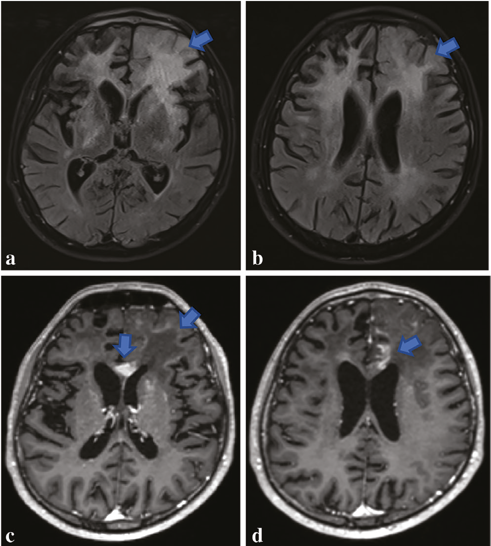
(a,b) Non-contrast brain MRI, January 2025: Axial FLAIR sequences demonstrate hyperintense lesions in the periventricular white matter of both frontal lobes and within the basal ganglia. A voluminous lesion involving the cortex and white matter is noted in the left frontal lobe, causing effacement of the adjacent cortical sulci (blue arrow). (c,d) Post-contrast T1-weighted images reveal peripheral linear enhancement in the anterior segment of the left frontal lesion, along with nodular and punctate enhancement at the level of the corpus callosum and the left caudate nucleus (blue arrows)
MRI: Magnetic resonance imaging, FLAIR: Fluid-attenuated inversion recovery
Within one month, the patient was readmitted because of worsening speech and increasing instability. A repeat brain MRI () demonstrated new contrast-enhancing lesions with diffusion restriction in the right hemisphere, while the size and enhancement of the previously noted left-sided lesions had decreased, likely reflecting a transient response to prior corticosteroid therapy. Consequently, digital subtraction angiography was performed to evaluate for vasculitis, and whole-exome sequencing (WES) was initiated. Digital subtraction angiography and WES results were unremarkable. A biopsy of the right frontal lobe was planned. The clinical materials and diagnostic data for this case report were obtained in February 2025.
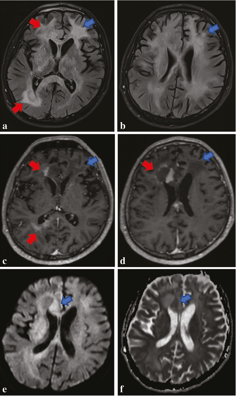
(a,b) Axial FLAIR sequence from February 2025 demonstrates regression in the size of the previously identified lesion in the anterior segment of the left frontal lobe (blue arrow). Concurrently, new lesions with significant mass effect and sulcal effacement have developed in the right frontal lobe adjacent to the corpus callosum (involving the cortico-medullary junction) and within the right periatrial white matter (red arrows). (c,d) Post-contrast images reveal ill-defined (faint) peripheral enhancement in the right frontal lesion and heterogeneous enhancement in the right periatrial white matter lesion (red arrows). The regressing lesion in the left frontal lobe shows no contrast enhancement (blue arrow). (e,f) Diffusion-weighted imaging shows hyperintensity along the medial aspect of the right frontal lesion, with corresponding mild hypointensity on the apparent diffusion coefficient map, indicating restricted diffusion. (a,d) In both January and February MRI studies, the cortical sulci were prominent, reflecting a decrease in brain parenchymal volume (atrophy) disproportionate to the patient’s age
MRI: Magnetic resonance imaging, FLAIR: Fluid-attenuated inversion recovery
Histopathological findings: Examination of sections from the right frontal lobe biopsy revealed an atypical lymphoid cell population infiltrating the brain parenchyma, characterized by blastic chromatin, prominent apoptosis, and increased mitotic activity (). Immunohistochemical analysis showed that the tumor cells were diffusely positive for CD20, PAX-5, Bcl-6, CD10, and c-MYC, while they were negative for Bcl-2, CD30, and ALK (). Within and adjacent to the tumor, a population of CD3-positive reactive T-lymphoid cells was observed, concentrating in perivascular areas and extending into the parenchyma ().
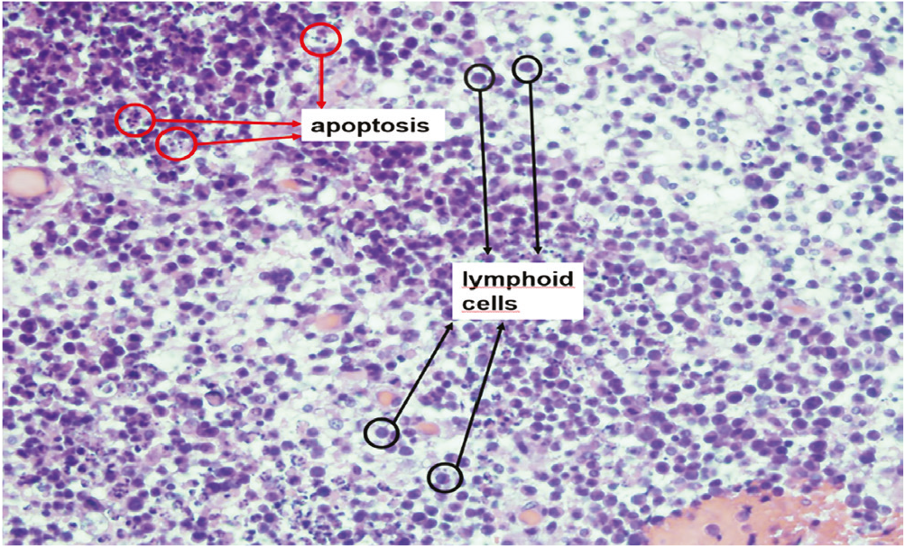
Histopathological examination demonstrates a monotonous population of medium-to-large lymphoid cells infiltrating the brain parenchyma. The cells exhibit blastic chromatin, scant cytoplasm, prominent apoptosis, and increased mitotic activity (H&E staining, ×400)
H&E: Hematoxylin and eosin
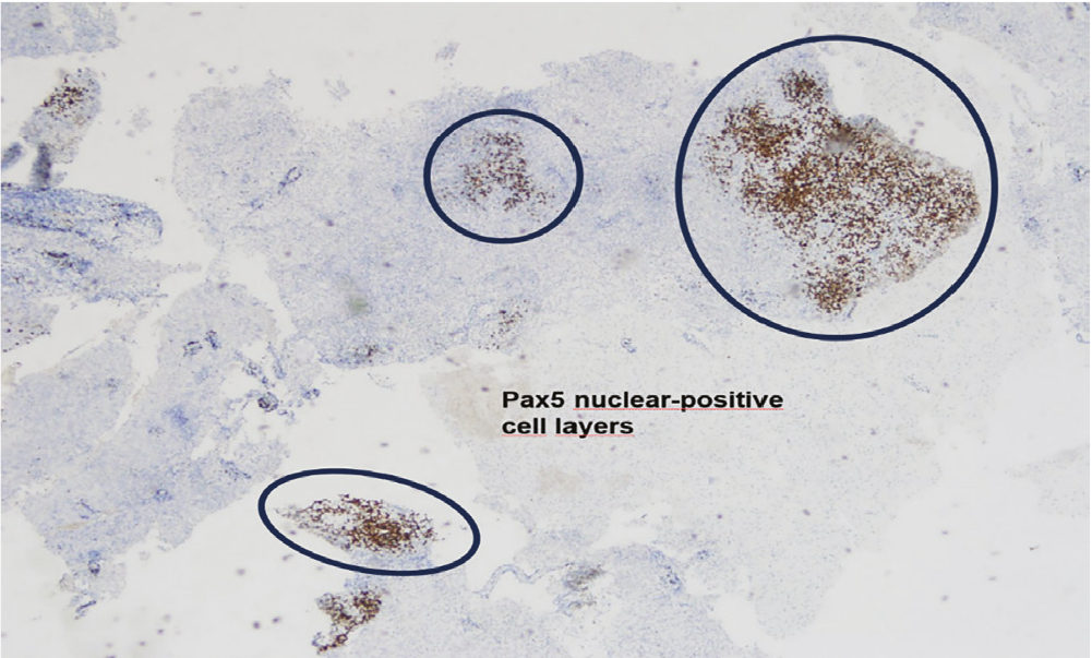
Photomicrograph showing diffuse and strong nuclear immunoreactivity for PAX5 within the neoplastic cells previously identified in Figure.3 (x40 magnification)
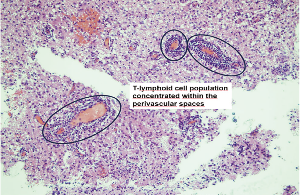
Photomicrograph demonstrates a T-lymphoid cell population concentrated within the perivascular spaces and extending into the brain parenchyma, observed both within the tumor and in the adjacent tissues (H&E, x200 magnification)
H&E: Hematoxylin and eosin
Discussion
Primary central nervous system lymphoma is an aggressive malignancy accounting for approximately 1-5% of all primary brain tumors and nearly 1% of all non-Hodgkin lymphomas (,). Although it predominantly affects immunocompromised individuals, its occurrence in immunocompetent young adults is rare and represents an atypical presentation that poses a significant clinical and radiological diagnostic challenge (,).
The differential diagnosis for PCNSL is notoriously broad, encompassing secondary lymphoma, glioblastoma, metastatic disease, viral or autoimmune encephalitis, and tumefactive demyelinating lesions (,). In the CNS, PCNSL primarily manifests as infiltrative parenchymal lesions. While multifocality and necrosis are more common among immunocompromised patients, immunocompetent patients often present with solitary, non-necrotic masses. Predilection sites include the periventricular white matter, basal ganglia, and corpus callosum (,). On MRI, PCNSL typically appears iso- to hypointense on both T1- and T2-weighted sequences, a key reflection of its high nuclear-to-cytoplasmic ratio and compact cellularity (,). A hallmark of the disease is marked diffusion restriction on apparent diffusion coefficient (ADC) maps, which serves as a critical differentiator from high-grade gliomas; PCNSL typically exhibits higher cellular density and lower ADC values than glial tumors (,). Following contrast administration, intense and homogeneous enhancement is the classic presentation, reflecting blood-brain barrier disruption ().
The radiological findings in this 18-year-old patient presented a significant diagnostic challenge, with an initial differential diagnosis encompassing viral or autoimmune encephalitis, tumefactive demyelinating lesions, and high-grade gliomas. The initial MRI, performed at an external facility in December 2024, revealed multifocal FLAIR hyperintensities involving the periventricular white matter and basal ganglia, suggesting an inflammatory or infectious process. However, the subsequent spatiotemporal evolution of the lesions provided the first critical clue for PCNSL. The regression of the left frontal lesion within one month, a classic manifestation of the “vanishing” or “ghost tumor” phenomenon, is highly characteristic of PCNSL’s sensitivity to corticosteroids or its naturally fluctuating course, a feature rarely seen in gliomas or metastatic disease (,). This transient response significantly hinders definitive diagnosis and delays essential treatment. Therefore, in cases where clinico-radiological discordance exists, early stereotactic biopsy is mandatory to circumvent diagnostic delays.
Furthermore, the emergence of new lesions in the right frontal and periatrial regions (February 2025) with significant mass effect and restricted diffusion (hyperintensity on diffusion-weighted imaging and mild hypointensity on ADC) served as a key radiologic differentiator. Restricted diffusion is a hallmark of the high cellularity (hypercellularity) typical of lymphoma, whereas autoimmune encephalitis typically lacks such focal restricted diffusion and rarely presents with the heterogeneous/peripheral enhancement patterns observed in this case (,). Additionally, while tumefactive demyelinating lesions may show peripheral enhancement, they typically exhibit an “open-ring” pattern and lack the dense, solid hypercellularity seen on ADC maps (,). Finally, the presence of brain atrophy disproportionate to the patient’s age added a layer of complexity, necessitating the exclusion of underlying systemic immunodeficiencies, which were subsequently ruled out, thereby confirming the atypicality of this PCNSL presentation in an immunocompetent young adult (,).
To contextualize our findings, summarizes recent literature on atypical PCNSL in young or immunocompetent patients. Unlike classic homogeneous masses, these cases increasingly exhibit atypical features such as the ‘vanishing tumor’ phenomenon or encephalitis-like presentation. This underscores the critical need to consider PCNSL in the differential diagnosis of atypical CNS lesions in young adults, regardless of immune status.
Table 1. Summary of recent literature highlighting atypical presentations and diagnostic challenges of Primary Central Nervous System Lymphoma (PCNSL)
Author, year (ref) | Study design | Main focus/atypical feature |
Van Der Meulen et al. (), 2026 | Review | Current standards and developments |
Ayalew et al. (), 2024 | Case report | Young immunocompetent patient |
Ribas et al. (), 2024 | Review | Imaging features and differential diagnosis |
Zawar et al. (), 2021 | Case report | Mimicking herpes encephalitis |
Histopathologically, while the majority of PCNSL cases are diffuse large B-cell lymphomas, our case presented a unique diagnostic conundrum (,). Initial immunohistochemical markers (specifically CD10 and Bcl-6 positivity and widespread c-MYC positivity) strongly raised suspicion for Burkitt lymphoma. However, a Ki-67 index of 85% and a prominent T-lymphoid population intermingled with the neoplastic cells were more consistent with high-grade large B-cell lymphoma. This dense, reactive cellular infiltrate, combined with the patient’s protracted clinical history, suggests a malignancy arising within an autoinflammatory background. Such a complex reactive environment likely acted as a histopathological masquerade, contributing to the initial diagnostic confusion and the “encephalitis-like” presentation that defined the early clinical course (). Ultimately, the absence of EBV-LMP1 and ALK expression further refined the diagnosis, confirming an aggressive PCNSL subtype in an immunocompetent host (,).
Conclusion
Primary central nervous system lymphoma is a rare and aggressive malignancy that can mimic glioblastoma, demyelinating lesions, encephalitis, and other neurological disorders, even in patients during the second decade of life. In patients presenting with atypical CNS lesions and negative viral, autoimmune, or metabolic workups, a fluctuating radiological pattern (characterized by the paradoxical regression of initial lesions and the emergence of new ones) should strongly heighten clinical suspicion for lymphoma. This case highlights the critical necessity of avoiding corticosteroid administration before a tissue biopsy. High-potency corticosteroids can induce rapid cytoreduction in PCNSL, leading to the “vanishing tumor” phenomenon, which may preclude a definitive histological diagnosis and delay life-saving treatment.
This case adds to the limited literature on PCNSL in young immunocompetent patients by emphasizing that a fluctuating radiological course (“vanishing tumor” phenomenon) may represent an important diagnostic clue. Early recognition of this pattern may shorten diagnostic delay, prevent inappropriate surgical management, and facilitate prompt initiation of lymphoma-directed therapy.
Ethics
Informed Consent: Written informed consent was obtained from the patient for the publication of this case report and any accompanying images.
Footnotes
Authorship Contributions
Concept: M.D.I., F.C., Design: M.D.I., F.C., Data Collection or Processing: M.D.I., D.T.S., F.C., Analysis or Interpretation: M.D.I., D.T.S., F.C., Literature Search: M.D.I., D.T.S., Writing: M.D.I., D.T.S., F.C.
Conflict of Interest
No conflicts of interest were declared by the authors.
Financial Disclosure
The authors declared that this study received no financial support.
References
Ferreri AJM, Calimeri T, Cwynarski K, et al. Primary central nervous system lymphoma. Nat Rev Dis Primers. 2023;9:29.
Van Der Meulen M, Vermaat JSP, Galldiks N, et al. Primary central nervous system lymphoma-current standards and recent developments. Neurooncol Pract. 2026;13:640-8.
Deckert M, Engert A, Brück W, et al. Modern concepts in the biology, diagnosis, differential diagnosis and treatment of primary central nervous system lymphoma. Leukemia. 2011;25:1797-807.
Ayalew ZS, Gebregiorgis M, Azibte GT, Hamza AK, Abdo IS, Molla BA. Primary central nervous system lymphoma: a diagnostic challenge in a young immunocompetent patient with limited resources. Radiol Case Rep. 2024;19:4644-9.
Han Y, Wang ZJ, Li WH, et al. Differentiation between primary central nervous system lymphoma and atypical glioblastoma based on MRI morphological feature and signal intensity ratio: a retrospective multicenter study. Front Oncol. 2022;12:811197.
Ribas GA, de Mori LH, Freddi TAL, Oliveira LDS, de Souza SR, Corrêa DG. Primary central nervous system lymphoma: imaging features and differential diagnosis. Neuroradiol J. 2024;37:705-22.
Haldorsen IS, Espeland A, Larsson EM. Central nervous system lymphoma: characteristic findings on traditional and advanced imaging. AJNR Am J Neuroradiol. 2011;32:984-92.
Slone HW, Blake JJ, Shah R, Guttikonda S, Bourekas EC. CT and MRI findings of intracranial lymphoma. AJR Am J Roentgenol. 2005;184:1679-85.
Feldheim J, Darkwah Oppong M, et al. Histopathological diagnosis of primary central nervous system lymphoma after therapy with corticosteroids or anticoagulants. Cancers (Basel). 2024;16:1157.
Zawar I, Cho SM, Prayson R, Levin K. Primary central nervous system lymphoma mimicking herpes encephalitis. Neurooncol Pract. 2021;8:620-2.