
Abstract
Aim
Pediatric ureteral stones present a clinical challenge because of anatomical limitations and the need for minimally invasive treatment approaches. The aim of this study was to assess outcomes of semi-rigid ureteroscopy (URS) in pediatric patients with mid- and distal-ureteral stones, with particular focus on predictors of postoperative stone-free (SF) status.
Methods
This single-center retrospective observational study included pediatric patients who underwent 4.5- and 6.5-Fr URS for mid- and distal-ureteral stones smaller than 2 cm between December 2016 and December 2024. Demographic characteristics, stone features, and perioperative outcomes were recorded. Participants were divided into two groups defined by the presence (n=17) or absence (n=52) of residual stones. Univariate and multivariate regression analyses were performed to identify factors associated with postoperative SF status.
Results
The study included 69 children (median age, 9 years; median stone size, 8 mm). Postoperative imaging revealed an SF rate of 75.4%. A history of surgery was less common in the SF group (p<0.001). The two groups differed in stone burden (p<0.001) and number of stones (p<0.001). Four (5.8%) patients had postoperative febrile urinary tract infections. Multivariate regression analysis revealed that stone burden (>10 mm vs. ≤10 mm) was the only predictor for residual fragments after URS for mid and distal ureteral stones (p=0.048).
Conclusion
Ureteroscopy is a safe, effective, and applicable minimally invasive treatment modality with low complication rates for ureteral stones in children. However, stone burden should be considered during patient selection and preoperative counseling, as it independently predicts postoperative SF status after ultrathin pediatric URS.
Keywords: Pediatric urolithiasis, ureteral stone, ureteroscopy, stone burden, stone-free status
Introduction
Urolithiasis represents a significant cause of morbidity across both adult and pediatric populations, exhibiting a globally escalating prevalence (,). Pediatric urolithiasis is characterized by a high recurrence rate, attributable to its frequent association with underlying metabolic, anatomical, and infectious etiologies (). Due to the high recurrence rate of pediatric urolithiasis and the likelihood of multiple surgical interventions, minimally invasive treatment is essential (). Achieving stone-free (SF) status remains a paramount objective in clinical management (). Treatment strategies are determined by a confluence of factors, including stone location, stone size, and the patient’s unique urinary tract anatomy.
Ureteral stones account for approximately 7% of all urinary calculi in children (). The management options for children presenting with ureteral stones include a trial of spontaneous passage, medical expulsive therapy (MET), semi-rigid ureteroscopy (URS), and extracorporeal shock wave lithotripsy. Pediatric ureteral stones below 5 mm frequently pass spontaneously, while larger stones usually require intervention. Shock wave lithotripsy is associated with limitations, notably reduced efficacy, the necessity for serial treatments and subsequent anesthetic events, and a resultant elevation in cumulative radiation dosage (). Ureteroscopy is often prioritized due to its expeditious achievement of stone clearance and minimization of patient discomfort (). Because URS is primarily used for mid and distal ureteral stones and treatment outcomes may vary according to stone location, we focused on this relatively homogeneous patient population. Excluding proximal ureteral stones allowed a more accurate evaluation of factors associated with postoperative SF status.
However, the existing literature on URS in pediatric patients is limited by several factors, including the lack of standardized surgical protocols, variability in instrument diameters, and heterogeneity in intracorporeal lithotripsy modalities. We hypothesized that ultrathin, semi-rigid URS would provide high SF rates and low complication rates in children with mid- and distal ureteral stones and that stone-related factors would be associated with postoperative SF status. In this context, we assessed the outcomes of 4.5/6.5 Fr (ultrathin) URS in children with mid- and distal-ureteral stones, focusing on predictors of postoperative SF status.
Materials and Methods
Compliance with Ethical Standards
A retrospective analysis was conducted on 69 pediatric patients who underwent unilateral URS between December 2016 and December 2024 for the treatment of mid- and distal ureteral stones measuring less than 2 cm. This single-center study was conducted in accordance with the Declaration of Helsinki. Ethical approval for the study was obtained from the Goztepe Prof. Dr. Suleyman Yalcin City Hospital Non-Interventional Clinical Research Ethics Committee (approval no.: 2025/0248, date: 23.10.2025).
Study Design
All patients underwent preoperative low-dose non-contrast computed tomography (NCCT). Patients with abnormal renal anatomy, proximal ureteral stones, pushback during surgery, ureteral stenosis, or incomplete medical records were excluded from the study. Given the established consensus that ureteral stones exceeding 5 mm typically necessitate endourological intervention, we included patients with such stones. Patients with preoperative stents were not included in the study. The patient selection process and the study cohort are summarized in the flow diagram ().
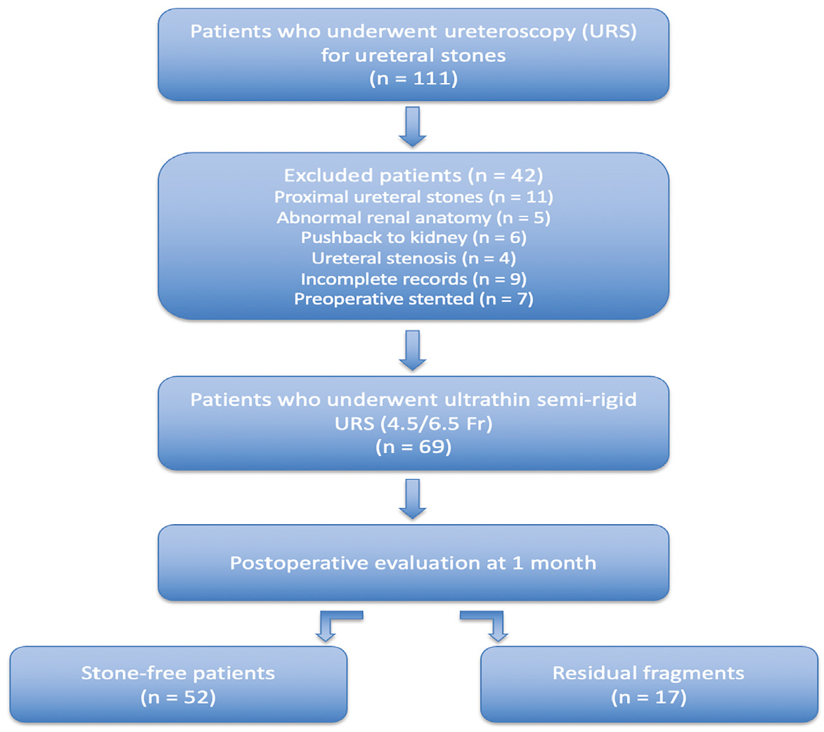
Study flow diagram showing patient selection and study cohort
Demographic data, degree of hydronephrosis, stone characteristics (laterality, number, location, size, and density), operative time, and perioperative and postoperative complications were recorded. Stone size was defined as the longest diameter of the stone, regardless of the NCCT axes. Stone density (Hounsfield unit) was quantified by manual delineation of the region of interest, which precisely conformed to the stone margins. In the case of multiple stones, stone burden was considered the sum of the stones’ longest diameters. The Society for Fetal Urology grading system was used to assess hydronephrosis severity on NCCT.
Operative Details
A negative urine culture was confirmed in all participants prior to the surgical intervention. All surgical procedures were conducted under general anesthesia. Prophylactic antibiotics were administered at a dose calculated according to guidelines. Case-specific surgical teams were assembled due to the long study period. However, all procedures were performed by urologists experienced in pediatric stone surgery, each of whom had performed at least 20 pediatric stone procedures before participating in the study. The same surgical principles and operative technique were applied throughout the study period. A 4.5/6.5 Fr semi-rigid ureteroscope (ultrathin; Richard Wolf GmbH, Knittlingen, Germany) served as the endoscopic instrument. Prior to the introduction of a safety guidewire (0.035-inch flexible) to the kidney, an examination of the urethra and bladder was performed. Fragmentation of the calculus was accomplished using a 272 µm Holmium:YAG laser fiber (Quanta System, Milan, Italy), with laser energy delivered in the range of 6 to 10 Watts. The dimensions of the stone fragments were assessed relative to the guidewire; fragments with a diameter equal to or smaller than that of the guidewire were left in situ. Stone fragments were irrigated with saline solution from the ureter to the bladder to promote spontaneous expulsion. After lithotripsy, a Foley catheter was inserted into all participants. The necessity and dimensions of double-J stent insertion were determined on an individualized basis for each patient. The urethral catheter was removed on the first postoperative day, and patients were subsequently discharged. In selected cases, extraction strings were used on the first postoperative day for stent removal.
Follow-up Procedure
At the first postoperative month, SF status was assessed by kidney-ureter-bladder (KUB) radiography and ultrasonography or by NCCT in cases of suspected residual calculus. To mitigate radiation exposure, NCCT was not routinely performed in all patients. Stone-free status was defined as the patient being completely SF or having residual fragments measuring less than 2 mm on radiological imaging. Double-J stents were removed within two weeks of the procedure in all patients, unless clinical indications necessitated extended stent retention. The primary endpoint of the study was postoperative SF status at the first-month follow-up.
Statistical Analysis
The data was analyzed using SPSS version 26 (Armonk, NY: IBM Corp., USA). Categorical variables in the study were presented with frequency (n) and percentage. For quantitative data, the Shapiro-Wilk test was used to assess the normality of the distribution. Quantitative variables with normal distribution were specified as mean ± standard deviation, and variables without normal distribution were presented as median (range) for descriptive statistics in the study. An independent samples t-test was used to compare the means. When the variables between groups were not normally distributed, the Mann-Whitney U test was used. For the analysis of categorical variables, the chi-squared test and Fisher’s exact test were used to compare the two groups. Univariate and multivariate logistic regression analyses were performed to determine the factors associated with residual fragments in the first month after surgery. Variables considered clinically relevant and those associated with residual fragments in the univariate analysis were included in the multivariable logistic regression model. Statistical significance was set at a p-value <0.05.
Results
Sixty-nine patients, comprising 46 boys and 23 girls, underwent URS. The median age was 9 years (range 1-17) and the median stone size was 8 mm (range 5-20). Postoperative imaging revealed an SF rate of 75.4% (n=52). Participants were divided into two groups based on SF status, and comparative analyses were performed. Statistical analysis revealed no significant differences between the groups regarding gender (p=0.429), age (p=0.710), family history (p=0.364), preoperative hydronephrosis (p=0.530), stone laterality (p=0.720), maximum stone size (p=0.187), stone density (p=0.928), or operation time (p=0.195). Surgical history was less common in the SF group (p<0.001). A difference between the two groups was observed for stone burden (p<0.001) and number of stones (p<0.001) ().
Table 1. Demographic and clinical characteristics of the groups
- | Stone-free (n=52) | Non-stone-free (n=17) | p-value |
|---|---|---|---|
Gender, n (%) Girl Boy | 16 (30.8) 36 (69.2) | 7 (41.2) 10 (58.8) | 0.429a |
Age (years), median (min-max) | 10 (1-17) | 7 (1-17) | 0.710b |
Family history, n (%) | 24 (46.1) | 10 (58.8) | 0.364a |
Surgical history, n (%) | 11 (21.1) | 14 (82.3) | <0.001a |
Preoperative hydronephrosis, n (%) None Grade 1 Grade 2 Grade 3-4 | 8 (15.4) 21 (40.4) 21 (40.4) 2 (3.8) | 2 (11.8) 4 (23.5) 10 (58.8) 1 (5.9) | 0.530a |
Laterality, n (%) Right Left | 24 (46.1) 28 (53.8) | 7 (41.2) 10 (58.8) | 0.720a |
Number of stones, n (%) Single Multiple | 48 (92.3) 4 (7.7) | 8 (47) 9 (53) | <0.001a |
Maximum stone size (mm), median (min-max) | 7 (5-20) | 8 (5-20) | 0.187b |
Stone burden (mm), median (min-max) | 8 (5-20) | 14 (5-28) | <0.001b |
Stone density (Hounsfield unit), median (min-max) | 762 (350-1735) | 900 (450-1700) | 0.928b |
Operation time (minutes), median (min-max) | 40 (15-90) | 45 (20-70) | 0.195b |
aPearson’s chi-squared test, bMann-Whitney U test | |||
The size of the ureteral stent was determined on a case-by-case basis; however, in 14 patients (20.3%), stent placement was not indicated due to the absence of clinical need. Within 24 hours of the procedure, 66 patients (95.6%) were discharged. Four patients (5.8%) had postoperative febrile urinary tract infection. Stone analysis was not feasible in 71% of participants (). An auxiliary URS procedure was required for 10 (14.5%) children who were not SF after the initial surgery.
Table 2. Postoperative clinical characteristics of participants
- | Participants (n=69) |
|---|---|
Ureteral stent placement, n (%) None 3 Fr Double-J stent 4 Fr Double-J stent 4.7 Fr Double-J stent | 14 (20.3) 16 (23.2) 13 (18.8) 26 (37.7) |
Postoperative febrile UTI, n (%) | 4 (5.8) |
Length of hospital stay, n (%) 1 day >1 day | 66 (95.6) 3 (4.3) |
Stone analysis, n (%) Not performed Calcium oxalate Cystine | 49 (71) 7 (10.1) 13 (18.8) |
UTI: Urinary tract infection | |
Multivariate regression analysis revealed stone burden (>10 mm vs. ≤10 mm) was the only predictor for residual fragments after URS for mid- and distal ureteral stones (p=0.048) ().
Table 3. Univariate and multivariate analysis: factors affecting residual fragments after URS
- | Univariate analysis | Multivariate analysis | ||
|---|---|---|---|---|
OR (95% CI) | p-value | OR (95% CI) | p-value | |
Age (>10 years vs. ≤10 years) | 0.809 (0.308-2.126) | 0.668 | - | - |
Surgical history (yes vs. no) | 8.213 (2.360-28.580) | 0.001 | 3.646 (0.899-14.782) | 0.070 |
Number of stones (multiple vs. single) | 4.846 (1.870-12.560) | 0.001 | 1.237 (0.414-3.702) | 0.703 |
Stone burden (>10 mm vs. ≤10 mm) | 9.333 (2.682-32.477) | <0.001 | 4.390 (0.979-19.691) | 0.048 |
Stone density (>800 HU vs. ≤800 HU) | 1.887 (0.698-5.103) | 0.211 | - | - |
Bold values indicate statistically significant p-values (<0.05) OR: Odds ratio, CI: Confidence interval, URS: Ureteroscopy, HU: Hounsfield unit
Discussion
Because the propensity for stone recurrence is higher in pediatric patients than in adults, the potential need for future surgical intervention should be considered, and minimally invasive techniques should be the initial therapeutic approach. The clinical management of pediatric urolithiasis is challenged by the inherent fragility and narrow diameter of the collecting system (). In spite of the challenges presented, technological innovations, specifically the miniaturization of endoscopic equipment, have improved the safety and efficacy of minimally invasive approaches in clinical practice (,). The development of ultrathin URS was undertaken to achieve a reduction in complication rates (). However, a recent survey showed that the availability of ultrathin URS is limited, with only 24% of 74 EAU centers having this equipment (). In this study, all cases were treated using our routinely used ultrathin URS, which has been available for pediatric stone procedures for many years. In our study, we used the Holmium:YAG laser for lithotripsy. While pneumatic lithotripters were previously common in pediatric URS, they are now rarely used due to higher complication rates and lower SF rates (,,).
The primary objective of URS for the management of ureteral calculi is the attainment of complete stone clearance. Published reports indicate URS success rates ranging from 75% to 100%, with superior outcomes observed for distal ureteral stones (,,-). Our SF rate (75.4%) is consistent with the existing literature. SF status was defined as the absence of fragments or the presence of fragments <2 mm, which is a stricter definition than that used in several previous pediatric urolithiasis studies (,). Our study cohort encompassed patients with a single ureteral calculus or multiple ureteral calculi. Consequently, we determined that stone burden affected SF status after ultrathin URS. The literature contains extensive evidence of a negative correlation between stone size and postoperative SF rates in children; however, studies evaluating the impact of stone burden on procedural success are limited (,,). Recent studies have identified ureteral wall thickness and ureteral diameter as potential predictors of SF status after URS (). As these parameters were not routinely recorded in our retrospective dataset, they could not be evaluated in the present study. Prospective studies may further clarify their role in predicting procedural success.
Ureteroscopy provides effective treatment with low morbidity and high patient satisfaction. Postoperative minor complications, including mucosal injury, hematuria, fever, and colic pain, may occur, with reported rates not exceeding 20% (,). Dogan et al. () reported an 8.4% complication rate in a retrospective, multi-institutional pediatric URS study, with operative time identified as the only factor affecting complication rates. In this study, laser use was limited to one out of five patients, and instruments larger than 8 Fr were used in more than half of the patients (). Our study demonstrated a low complication rate: transient fever was the only observed adverse event, affecting four patients (5.8%). A 95.6% next-day discharge rate was achieved, attributable to a remarkably low complication profile. The use of small-caliber URS and the limited duration of the procedures are hypothesized to contribute to this favorable outcome. In our study, pushback to the kidney was not observed; this finding is attributable to the absence of proximal ureteral calculi. Furthermore, major complications such as perforation, ureteral rupture, or sepsis, which have been documented in the literature, did not occur in our series ().
In our study, active dilation of the ureteral orifice was intentionally omitted in all patients due to the potential for complications, including ureteral stenosis and vesicoureteral reflux (). Two recent investigations have reported access failure rates of 16.6% and 20% when employing 6 Fr and 7.5 Fr semi-rigid ureteroscopes in pediatric patients, respectively (,). In concordance with findings reported in previous studies, ultrathin URS facilitated successful ureteral access in all patients, notwithstanding the notably younger age distribution of our cohort (,). Postoperative stenting remains a subject of debate. However, it is believed to promote enhanced healing and resolution of local edema, while also mitigating postoperative pain and the risk of infectious complications arising from obstruction (). Ureteral stenting after pediatric URS is reported in 31-100% of cases (). However, stent removal necessitates an additional procedure under general anesthesia. Given the relatively young age of our patient cohort and the potential for postoperative renal colic secondary to mucosal edema, temporary catheter placement was performed in 55 of 69 patients. In addition, despite the demonstrated efficacy of MET in children, its off-label status and potential safety concerns precluded its use in our study (,,).
Study Limitations
Our study has limitations, including its retrospective design. Additionally, we primarily used KUB radiography and ultrasonography instead of NCCT to assess SF status to reduce radiation exposure in children; this choice may have influenced the reported SF rate. The lower SF rate observed in our cohort, compared with some reports in the literature, may also be explained by the inclusion of patients with multiple stones and a higher overall stone burden. Other significant limitations include the limited number of patients and the wide age range. In addition, the relatively small number of residual stone events (n=17) may have increased the risk of overfitting in the multivariable logistic regression model. Therefore, the identified predictors should be interpreted with caution and validated in larger prospective cohorts. Stone analysis was not available for all patients because of our predominant use of the dusting technique. Furthermore, due to limitations inherent in the retrospective dataset, a subgroup analysis comparing SF outcomes between mid and distal ureteral stones could not be performed. While the same surgical technique was consistently employed by all surgeons within this cohort, the possibility of inter-surgeon differences affecting both success and complication rates remains. This factor should be acknowledged as a limitation that influences the demonstrative capacity of the data. Despite these limitations, this study makes a significant contribution to the literature as one of the largest series validating the safety and efficacy of ultrathin URS for pediatric ureteral stone management.
Conclusion
Ureteroscopy is a safe, effective, and feasible minimally invasive treatment modality with low complication rates for ureteral stones in children. Our study demonstrates that URS in children yields favorable outcomes, characterized by short hospital stays and high SF rates. The success of the procedure is significantly affected by the stone burden. Therefore, stone burden should be considered during patient selection and preoperative counseling because it independently predicts postoperative SF status after ultrathin pediatric URS.
Ethics
Ethics Committee Approval: The ethics committee approved the commencement of the present study Goztepe Prof. Dr. Suleyman Yalcin City Hospital Non-Interventional Clinical Research Ethics Committee (approval no.: 2025/0248, date: 23.10.2025). All authors fully complied with the Declaration of Helsinki during the course of the study.
Informed Consent: The ethics committee waived the requirement for informed consent because the study was retrospective.
Footnotes
Authorship Contributions
Surgical and Medical Practices: A.I., M.C., O.A., M.S.S., Concept: A.I., M.C., A.Y., Design: A.I., M.C., O.A., M.S.S., Data Collection or Processing: A.I., M.C., M.K., Analysis or Interpretation: A.I., O.A., A.Y., Literature Search: A.I., O.A., Writing: A.I.
Conflict of Interest
No conflicts of interest were declared by the authors.
Financial Disclosure
This study received no financial support.
References
Sorokin I, Mamoulakis C, Miyazawa K, Rodgers A, Talati J, Lotan Y. Epidemiology of stone disease across the world. World J Urol. 2017;35:1301-20.
Utangaç MM, Dede O. Evaluation of the largest series of ultramini percutaneous nephrolithotomy in preschool children: 10-year experience with 711 units of kidney stones. J Clin Med. 2025;14:3355.
Gajengi AK, Wagaskar VG, Tanwar HV, Mhaske S, Patwardhan SK. Metabolic evaluation in paediatric urolithiasis: a 4-year open prospective study. J Clin Diagn Res. 2016;10:PC04-6.
Baştuğ F, Gündüz Z, Tülpar S, Poyrazoğlu H, Düşünsel R. Urolithiasis in infants: evaluation of risk factors. World J Urol. 2013;31:1117-22.
Chew BH, Brotherhood HL, Sur RL, et al. Natural history, complications and re-intervention rates of asymptomatic residual stone fragments after ureteroscopy: a report from the EDGE research consortium. J Urol. 2016;195:982-6.
Raza A, Smith G, Moussa SA, Tolley DA. Ureteroscopy in the management of pediatric urinary tract calculi. J Endourol. 2005;19:151-8.
Smaldone MC, Cannon GM, Wu H-Y, et al. Is ureteroscopy first line treatment for pediatric stone disease?. J Urol. 2007;178:2128-31.
Radmayr C, Bogaert G, Bujons A et al. EAU Guidelines on Paediatric Urology. 2026. Available from: https://d56bochluxqnz.cloudfront.net/documents/full-guideline/EAU-Guidelines-on-Paediatric-Urology-2026.pdf
Yucel S, Akın Y, Kol A, Danışman A, Guntekin E. Experience on semirigid ureteroscopy and pneumatic lithotripsy in children at a single center. World J Urol. 2011;29:719-23.
Ishii H, Griffin S, Somani BK. Ureteroscopy for stone disease in the paediatric population: a systematic review. BJU Int. 2015;115:867-73.
Onal B, Citgez S, Tansu N, et al. What changed in the management of pediatric stones after the introduction of minimally invasive procedures? A single-center experience over 24 years. J Pediatr Urol. 2013;9:910-4.
Herndon CD, Viamonte L, Joseph DB. Ureteroscopy in children: is there a need for ureteral dilation and postoperative stenting?. J Pediatr Urol. 2006;2:290-3.
Bañuelos Marco B, Haid B, Radford A, et al. Guideline adherence of paediatric urolithiasis: an EAU Members’ Survey and expert panel roundtable discussion. Children (Basel). 2022;9:504.
Irsayanto D, Setiawan MR, Salsabila S, et al. Efficacy and safety of pneumatic lithotripsy with laser lithotripsy in the treatment of ureteral stones <20 millimeters in children: a systematic review and meta-analysis. J Ayub Med Coll Abbottabad. 2024;36:201-8.
Turunc T, Kuzgunbay B, Gul U, et al. Factors affecting the success of ureteroscopy in management of ureteral stone diseases in children. J Endourol. 2010;24:1273-7.
Utanğaç MM, Sancaktutar AA, Tepeler A. Micro-ureteroscopy for the treatment of distal ureteral calculi in children. J Pediatr Surg. 2017;52:512-6.
Dogan HS, Tekgul S, Akdogan B, Keskin MS, Sahin A. Use of the Holmium:YAG laser for ureterolithotripsy in children. BJU Int. 2004;94:131-3.
Sinha M, Pietropaolo A, Quiroz Madarriaga Y, et al. Outcomes of ureteroscopy for management of stone disease in early and late childhood over a 15-year period. Ther Adv Urol. 2022;14:17562872221141775.
Amer ML, Elkholefy ME, Dawoud H, Gaber M, El-Gamal OM, Gameel T. Safety and efficacy of Silodosin as medical expulsive therapy after shock wave lithotripsy in paediatric patients with renal stones. Urolithiasis. 2025;53:95.
Ahmed A, Farhan B, Alam-Eldin A, et al. A prospective randomized trial comparing dusting and fragmentation techniques using Holmium:YAG laser for pediatric ureteral stones. Can J Urol. 2025;32:483-90.
Jaidane M, Hidoussi A, Slama A, Hmida W, Ben Sorba N, Mosbah F. Factors affecting the outcome of ureteroscopy in the management of ureteral stones in children. Pediatr Surg Int. 2010;26:501-4.
Kucukdurmaz F, Efe E, Sahinkanat T, Amasyalı AS, Resim S. Ureteroscopy with Holmium:YAG laser lithotripsy for ureteral stones in preschool children: analysis of the factors affecting the complications and success. Urology. 2018;111:162-7.
Selvi I, Baydilli N, Tokmak TT, Akinsal EC, Basar H. CT-related parameters and Framingham score as predictors of spontaneous passage of ureteral stones ≤ 10 mm: results from a prospective, observational, multicenter study. Urolithiasis. 2021;49:227-37.
Dogan HS, Onal B, Satar N, et al. Factors affecting complication rates of ureteroscopic lithotripsy in children: results of multi-institutional retrospective analysis by Pediatric Stone Disease Study Group of Turkish Pediatric Urology Society. J Urol. 2011;186:1035-40.
Topaktas R, Aydin C, Altin S, Akkoc A, Aydın ZB, Urkmez A. The efficacy of ultra-thin semi-rigid ureteroscopy with holmium laser lithotripsy in pediatric ureteral stones: a single-center experience. Cureus. 2019;11:e5496.
Tiryaki T, Azili MN, Özmert S. Ureteroscopy for treatment of ureteral stones in children: factors influencing the outcome. Urology. 2013;81:1047-51.
Hubert KC, Palmer JS. Passive dilation by ureteral stenting before ureteroscopy: eliminating the need for active dilation. J Urol. 2005;174:1079-80; discussion 1080.
AbdelRazek M, Fathi A, Mohamed M, Abdel-Kader MS. Primary versus deferred ureteroscopy for the management of obstructive anuria secondary to ureteric urolithiasis in children: a prospective randomized study. Urolithiasis. 2022;51:6.
Atar M, Sancaktutar AA, Penbegul N, et al. Comparison of a 4.5 F semi-rigid ureteroscope with a 7.5 F rigid ureteroscope in the treatment of ureteral stones in preschool-age children. Urol Res. 2012;40:733-8.
Kocaoglu C, Ozkan KU. The effectiveness of 4.5F ultrathin semirigid ureteroscope in the management of ureteral stones in prepubertal children: is there a need for any ureteral dilatation?. Urology. 2014;84:202-5.
Mokhless I, Zahran AR, Youssif M, Fahmy A. Tamsulosin for the management of distal ureteral stones in children: a prospective randomized study. J Pediatr Urol. 2012;8:544-8.
Elgalaly H, Eliwa A, Seleem M, et al. Silodosin in the treatment of distal ureteric stones in children: a prospective, randomised, placebo-controlled study. Arab J Urol. 2017;15:194-8.