
Abstract
Aim
Peptidylarginine deiminase type 4 (PADI4), a key enzyme involved in neutrophil extracellular trap (NET) formation, has been implicated in the pathogenesis of autoinflammatory diseases, including Behçet’s disease (BD). We aimed to evaluate serum PADI4 levels in neuro-BD (NBD) and to investigate their association with disease activity and clinical parameters.
Methods
This study was designed as an analytical cross-sectional study between 2021 and 2023. A total of 25 patients with NBD, 45 with multiple sclerosis, 10 with neuromyelitis optica (NMO), and 19 healthy controls were enrolled. Serum PADI4 concentrations were measured using ELISA. Comprehensive neuropsychological assessments were conducted in the NBD group. Correlation analyses were performed to evaluate associations with cognitive and clinical parameters.
Results
Serum PADI4 levels were significantly elevated in NBD patients compared to healthy controls but were comparable among patient groups. None of the cognitive and motor performance scores were correlated with PADI4 levels. NBD patients with a positive pathergy test showed significantly higher PADI4 levels.
Conclusion
Neutrophil extracellular traposis may represent a shared inflammatory pathway across immune-mediated brain disorders. However, the lack of association between serum PADI4 levels and clinical or cognitive parameters in NBD suggests limited diagnostic and prognostic utility as a biomarker.
Keywords: Peptidylarginine deiminase type 4, neutrophil extracellular trap, autoinflammatory disease, biomarker, Behçet’s disease
Introduction
Neuro-Behçet’s disease (NBD), an uncommon manifestation of Behçet’s disease (BD), is an inflammatory disorder characterized by recurrent and unpredictable relapsing-remitting episodes of neurological involvement occurring in approximately 5-10% of individuals diagnosed with BD (). According to magnetic resonance imaging (MRI) findings, NBD can be classified as parenchymal or non-parenchymal and may lead to neurocognitive impairments, particularly in attention, delayed recall, working memory, and executive functioning (, ). BD is considered to have predominantly autoinflammatory features, involving dysregulated neutrophil activity (). Neutrophils contribute to inflammation by generating neutrophil extracellular traps (NETs), which are released through a mechanism called NETosis and accumulate in serum and inflamed tissues, thereby promoting endothelial damage and a procoagulant state (). The pathergy test, a diagnostic tool for BD, is characterized by non-specific skin hypersensitivity and intensive neutrophil infiltration, which clinically mirrors this hyperinflammatory profile ().
Peptidylarginine deiminase type 4 (PADI4), a calcium ion-dependent enzyme, catalyzes protein citrullination by converting arginine to citrulline, thereby altering protein structure and function (). It plays a critical role in inflammatory processes by mediating histone citrullination during NET formation and has been associated with many autoimmune conditions due to its effects on immune cell dysregulation (). In multiple sclerosis (MS), PADI4 significantly facilitates the citrullination of myelin basic protein, leading to immune-mediated tissue damage. The direct association of PADI4 with BD remains unconfirmed; however, its role in critical neuro-inflammatory and autoimmune pathways emphasizes its potential relevance in the disease’s pathophysiology (). Therefore, we hypothesized that PADI4-mediated citrullination and NETosis lead to neuroinflammatory damage in NBD, and that PADI4 levels might correlate with disease activity.
This study aimed to investigate the role of PADI4 in NBD, with a particular focus on its involvement in immune-inflammatory processes and its potential association with disease activity. Serum PADI4 levels were compared among patients with NBD, other inflammatory brain disorders, and healthy controls, and their relationships with clinical and cognitive parameters were evaluated.
Materials and Methods
Compliance with Ethical Standards
Ethical approval was taken from the Istanbul University Faculty of Medicine Clinical Research Ethics Committee (approval no: 09, date: 16.04.2021). The study protocol was approved by the Institutional Review Board. The study was conducted in accordance with the ethical principles of the Declaration of Helsinki. All subjects provided written informed consent prior to any study-related procedure.
Study Design
This study was designed as an analytical cross-sectional study between 2021 and 2023. A total of 25 patients diagnosed with NBD according to the International Criteria for BD were enrolled in the study [median: 7.30 (interquartile ranges (IQR): 2.13); 14 men]. Additionally, 45 age- and gender-matched patients with [relapsing-remitting MS (RRMS), median: 6.48 (IQR: 2.92), 21 men], diagnosed according to the revised McDonald criteria, 10 patients with [neuromyelitis optica (NMO), median: 6.35 (IQR: 2.02), 5 men], and 19 healthy controls [healthy control, median: 3.58 (IQR: 1.97), 10 men] were included as control groups. Due to the exploratory nature of the study, a formal sample size calculation was not performed; however, all eligible patients during the study period were included ().
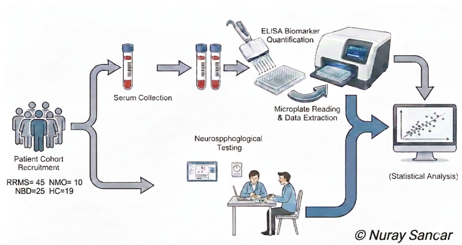
RRMS: Relapsing-remitting multiple sclerosis, NMO: Neuromyelitis optica, NBD: Neuro-Behçet’s disease, HC: Healthy controls, ELISA: Enzyme-linked immunosorbent assay
Serum samples were obtained from all NBD patients and controls during remission and stored at -80 °C until use. Neuropsychological tests were administered during serum sampling. Magnetic resonance imaging and magnetic resonance venography confirmed typical parenchymal NBD findings—such as unilateral or bilateral hyperintense lesions extending from the brainstem to the diencephalon and basal ganglia—and showed no cerebral venous thrombosis indicative of non-parenchymal involvement.
ELISA
Human PADI4 levels were measured in serum samples using an ELISA kit (Bioassay Technology Laboratory, China) according to the manufacturer’s instructions. Optical densities were measured at 450 nm, and concentrations were calculated based on standard curves provided in the kit. The results were expressed as ng/mL.
Neuropsychological Evaluation
General Cognitive Performance: The Mini-Mental State Examination and the Addenbrooke’s Cognitive Examination-Revised (ACE-R) were administered to evaluate overall cognitive capacity.
Attention and Orientation: The ACE-R Attention and Orientation subtest and the Wechsler Memory Scale-Revised (WMS-R) Digit Span (forward and backward) were used to assess attention and working memory performance.
Memory The ACE-R Memory subtest, WMS-R Visual Memory (immediate recall, long-term recall, and recognition), and the California Verbal Learning Test-II were used to evaluate verbal and visual memory performance.
Language skills were assessed using the ACE-R Language subtest and the Boston Naming Test (spontaneous, semantic cue, phonemic cue, paraphasia, and functional knowledge).
The Clock Drawing Test, Stroop Test (interference times, errors, and spontaneous corrections), Trail Making Test (TMT) A and B (completion time and errors), Wisconsin Card Sorting Test, the Symbol Digit Modalities Test, and the Luria Perseveration Test were administered to assess executive functions.
The TMT A and B, the 9-Hole Peg Test (placement and removal times for dominant and non-dominant hands), and the Timed 25-Foot Walk Test were administered to evaluate psychomotor speed and mental flexibility.
Verbal and categorical fluency were assessed using the ACE-R Fluency subtest, the (Controlled Oral Word Association Test; animal category, perseverations, and out-of-category words), the (Category Fluency Score; total score, perseverations, out-of-category words, and proper names), and the KAS Total Score.
Visuospatial abilities were examined using the ACE-R Visuospatial subtest, the Benton Facial Recognition Test, the Benton Line Orientation Test, and the Figure Copying Test.
Statistical Analysis
Statistical analyses were performed using GraphPad Prism software. Data distribution was assessed using the Kolmogorov-Smirnov test and found to be non-normal; therefore, results are presented as median (interquartile range). Comparisons of serum PADI4 levels among NBD, RRMS, NMO, and healthy control groups were conducted using the Kruskal-Wallis test, followed by Dunn-Bonferroni post-hoc analysis. Pairwise comparisons were performed using the Mann-Whitney U test. Correlations between PADI4 levels and demographic, clinical, and neuropsychological parameters were evaluated using Spearman’s rank correlation coefficient. A two-tailed p-value <0.05 was considered statistically significant, and Bonferroni correction was applied for multiple comparisons.
Results
Demographic and Clinical Characteristics of the NBD Group
The NBD group consisted of 25 patients (14 men and 11 women). The median duration of BD was 13 (IQR: 15), while the median duration of neurological involvement was 13 (IQR: 15). Pathergy testing was positive in 6 patients and negative in 13. Brain MRI findings revealed brainstem lesions in 15 patients, diencephalic involvement in 9 patients, and spinal cord lesions in 1 patient. The Expanded Disability Status Scale (EDSS) score had a median of 2 (IQR: 1.75). Detailed clinical and radiological characteristics of the NBD patients are presented in .
Table 1. Clinical, demographic features of NBD patients and controls
Feature | RRMS | NMO | NBD | HC |
|---|---|---|---|---|
Age (years) | Median: 52.0 (IQR: 9.5) | Median: 49.5 (IQR: 11.0) | Median: 52.0 (IQR: 4.0) | Median: 51.50 (IQR: 9.75) |
Gender (men/women) | 21/24 | 5/5 | 14/11 | 10/9 |
BD duration (years) | NA | NA | Median: 22 (IQR:10) | NA |
NBD duration (years) | NA | NA | Median: 13 (IQR:15) | NA |
Pathergy test (positive/negative)* | NA | NA | 6/13 | NA |
MRI lesions (brainstem) | ND | ND | 15 patients | NA |
MRI lesions (diencephalon) | ND | ND | 9 patients | NA |
MRI lesions (spinal cord) | ND | ND | 1 patient | NA |
EDSS | ND | ND | Median: 2 (IQR:1.75) | NA |
*Note that the pathergy test was available for 19 patients. Continuous variables are presented as median (interquartile range).
RRMS: Relapsing remitting multiple sclerosis, NMO: Neuromyelitis optica, HC: Healthy control, ND: Not determined, NA: Not applicable, IQR: Interquartile range
Comparison of Serum PADI4 Levels Among NBD, RRMS, NMO, and Healthy Controls
The Kruskal-Wallis test yielded a significant p-value for multiple group comparisons of all groups (p<0.0001). Dunn’s post-hoc test for multiple comparisons showed that NBD patients had significantly higher PADI4 levels than healthy controls (p<0.0001). Furthermore, both RRMS (p<0.0001) and NMO (p<0.0001) groups had significantly elevated levels of PADI4 compared with healthy controls. There were no differences among NBD, RRMS, and NMO groups in serum PADI4 levels. The graphical representation of these comparisons was provided in .
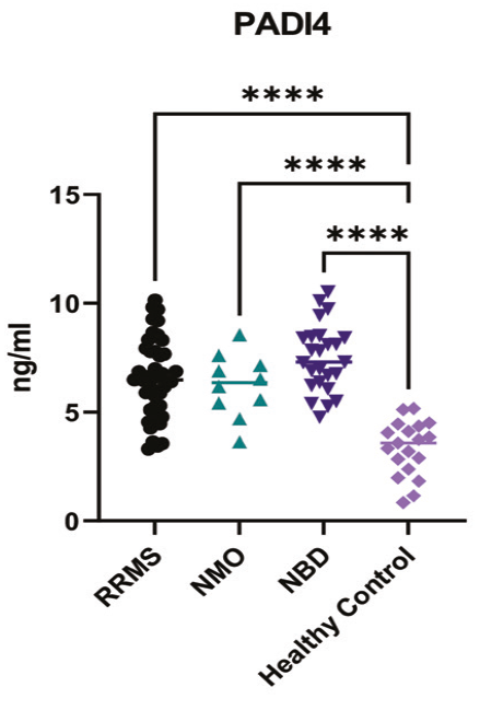
RRMS: Relapsing-remitting multiple sclerosis, NMO: Neuromyelitis optica, NBD: Neuro-Behçet’s disease, PADI4: Peptidylarginine deiminase type 4
Correlation Between PADI4 Levels and Clinical/Cognitive Features in NBD
No significant correlations were found between PADI4 levels and age, disease duration, and EDSS score (p>0.05 for all).
To explore the potential association between serum PADI4 levels and cognitive performance in patients with NBD, a series of neuropsychological and motor performance tests was administered. These tests assessed a wide range of cognitive domains, including general cognitive status, memory, language, executive function, attention, visuospatial skills, and psychomotor speed. Median scores, IQR, and correlation p-values between PADI4 levels and individual test results were presented in .
Table 2. Neuropsychological test scores of NBD patients: median, interquartile range (IQR), p and r values of correlation tests with PADI4 levels
Test name | Median | IQR | p-value | r-value |
|---|---|---|---|---|
MMSE/max 30 | 25.0 | 7.0 | 0.517 | 0.142 |
ACE-R total/max 100 | 69.0 | 19.0 | 0.442 | 0.168 |
ACE-R attention and orientation/max 18 | 16.0 | 6.0 | 0.478 | 0.155 |
ACE-R memory/max 26 | 12.0 | 7.0 | 0.662 | 0.096 |
ACE-R fluency/max 14 | 10.0 | 4.0 | 0.446 | 0.167 |
ACE-R language/max 26 | 20.0 | 9.0 | 0.314 | 0.207 |
ACE-R visuospatial/max 16 | 3.0 | 3.0 | 0.297 | 0.226 |
WMS-R digit span (forward) | 5.0 | 0.5 | 0.473 | 0.186 |
WMS-R digit span (backward) | 4.0 | 1.0 | 0.966 | 0.010 |
WMS-R visual memory (immediate) | 7.0 | 6.0 | 0.381 | 0.226 |
WMS-R visual memory (long-term recall) | 5.0 | 4.5 | 0.950 | 0.016 |
WMS-R visual memory (long-term recognition) | 2.0 | 1.5 | 0.468 | 0.188 |
Verbal fluency (fruit-name) | 6.5 | 5.0 | 0.375 | 0.229 |
COWAT animal score | 16.0 | 7.5 | 0.268 | 0.284 |
COWAT animal perseveration | 0.6 | 1.0 | 0.665 | -0.113 |
KAS total score | 19.0 | 23.0 | 0.740 | -0.086 |
KAS perseveration | 1.0 | 1.5 | 0.456 | -0.193 |
KAS out of category | 0.1 | 0.3 | 0.556 | -0.153 |
KAS proper name | 0.8 | 1.0 | 0.735 | 0.088 |
Stroop interference time | 66.5 | 41.5 | 0.853 | -0.054 |
Stroop spontaneous correction | 2.5 | 3.25 | 0.559 | -0.170 |
Stroop error | 3.50 | 9.25 | 0.451 | 0.219 |
Trail making test A - time | 82.0 | 42.75 | 0.969 | 0.010 |
Trail making test A - errors | 0.2 | 0.4 | 0.542 | -0.164 |
Trail making test B - time | 255 | 132.5 | 0.716 | -0.111 |
Trail making test B - errors | 2.0 | 2.0 | 0.160 | 0.413 |
Clock drawing test | 5.0 | 2.0 | 0.143 | 0.370 |
Luria perseveration (present:1 absent: 0) | 0.4 | 1.0 | 0.756 | -0.081 |
Symbol digit modalities test (SDMT) total items | 21.0 | 14.5 | 0.202 | 0.336 |
Symbol digit modalities test total correct | 20.5 | 16.5 | 0.261 | 0.298 |
Figure copying test | 3.0 | 2.0 | 0.444 | 0.198 |
CVLT II total correct | 33.0 | 16.5 | 0.980 | -0.006 |
CVLT II B list | 4.0 | 2.5 | 0.552 | -0.155 |
CVLT II short-term cued recall | 6.0 | 5.0 | 0.450 | -0.196 |
CVLT II long-term cued recall | 7.0 | 4.5 | 0.951 | 0.016 |
CVLT II short-term uncued recall | 4.0 | 5.5 | 0.711 | 0.096 |
CVLT II long-term uncued recall | 7.0 | 5.0 | 0.358 | -0.237 |
CVLT II total | 9.0 | 12.0 | 0.453 | -0.195 |
CVLT II recognition | 13.0 | 6.5 | 0.422 | 0.208 |
CVLT II recognition (YP) | 4.0 | 5.5 | 0.346 | -0.243 |
Boston naming test - spontaneous | 23.0 | 8.0 | 0.323 | 0.255 |
Boston naming test - no naming | 1.0 | 7.5 | 0.322 | -0.255 |
Boston naming test - semantic cue | 2.0 | 2.0 | 0.023 | -0.544 |
Boston naming test - phonemic cue | 2.0 | 3.0 | 0.596 | 0.138 |
Boston naming test - function | 0.5 | 0.9 | 0.105 | 0.406 |
Boston naming test - paraphasia | 0.0 | 0.5 | 0.215 | 0.316 |
Benton facial recognition test | 43.0 | 9.0 | 0.437 | 0.201 |
Benton line orientation test | 17.5 | 6.5 | 0.458 | 0.199 |
Wisconsin card sorting test (WCST) total responses | 125.5 | 34 | 0.981 | 0.006 |
Wisconsin card sorting test total errors | 40.0 | 35.25 | 0.850 | -0.051 |
Wisconsin card sorting test total correct | 74.5 | 27.0 | 0.832 | 0.057 |
Wisconsin card sorting test categories completed | 5.0 | 4.0 | 0.103 | 0.422 |
Wisconsin card sorting test perseverative responses | 22.5 | 25.5 | 0.930 | -0.023 |
Wisconsin card sorting test perseverative errors | 20.0 | 21.75 | 0.956 | -0.014 |
Wisconsin card sorting test non-perseverative errors | 14.0 | 14.0 | 0.665 | -0.121 |
Wisconsin card sorting test perseverative error percentage | 17.09 | 16.75 | 0.903 | 0.033 |
Wisconsin card sorting test first category completion trials | 13.0 | 9.0 | 0.786 | 0.076 |
Wisconsin card sorting test conceptual level responses | 68.0 | 34.75 | 0.748 | 0.087 |
Wisconsin card sorting test conceptual level response percentage | 53.45 | 31.68 | 0.557 | 0.158 |
Failure to maintain set score | 1.0 | 2.0 | 0.059 | -0.481 |
9-hole peg test placement (dominant hand) | 16.0 | 6.25 | 0.071 | -0.448 |
9-hole peg test placement (non-dominant hand) | 15.5 | 7.58 | 0.106 | -0.404 |
9-hole peg test removal (dominant hand) | 9.0 | 4.56 | 0.066 | -0.454 |
9-hole peg test removal (non-dominant hand) | 8.5 | 4.75 | 0.046 | -0.488 |
25-foot walk test | 16.0 | 11.75 | 0.967 | -0.010 |
Spearman correlation test was used for determination of p-values
MMSE: Mini-mental state examination, ACE-R : Addenbrooke’s Cognitive Examination-Revised, WMS-R: Wechsler Memory Scale-Revised, COWAT: Controlled Oral Word Association Test, CVLT: California Verbal Learning Test, NBD: Neuro-Behçet’s disease, PADI4: Peptidylarginine deiminase type 4
Overall, correlation analyses revealed no statistically significant associations between PADI4 levels and neuropsychological test scores. However, weak but statistically significant correlations were observed between PADI4 levels and the Boston Naming Test–Semantic Cue condition (p=0.023) and the 9-Hole Peg Test (Non-Dominant Hand–Removal Time) (p=0.046). However, these p-values did not remain significant after Bonferroni correction, which required p-values less than 0.00001.
Evaluation of Pathergy Test Results and Association with PADI4 Levels in NBD
To assess the relationship between inflammatory response markers and immune hyperreactivity, PADI4 levels were compared between pathergy-positive and pathergy-negative NBD patients. Using the Mann-Whitney U test, a statistically significant difference in serum PADI4 levels was found between the two groups (p=0.046, two-tailed, exact p-value). Patients who were pathergy-positive exhibited higher median PADI4 levels (median =8.434 ng/mL, n=6) compared to those who were pathergy-negative (median =6.91 ng/mL, n=13). The graphical representation of this comparison is shown in .
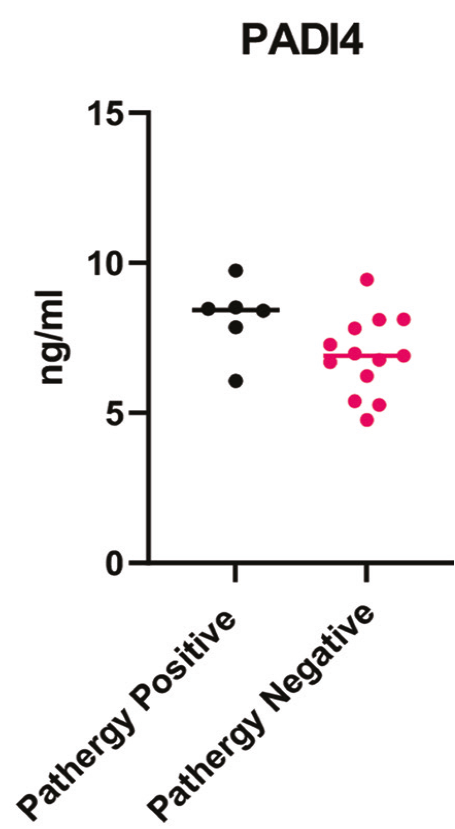
The horizontal line indicates median values. The p-value (p=0.046) was obtained by the Mann-Whitney U test
PADI4: Peptidylarginine deiminase type 4, NBD: Neuro-Behçet’s disease
Discussion
To our knowledge, this study provides additional evidence that serum PADI4 levels are elevated in patients with NBD compared to healthy controls. Furthermore, higher PADI4 levels were observed in patients with positive pathergy tests. Thus, our study provides new evidence supporting the role of PADI4 in the immunopathology of NBD. On the other hand, our study and previous studies have also shown that PADI4 elevation is not specific to NBD or BD and may also be observed in other immune-mediated disorders such as systemic lupus erythematosus, rheumatoid arthritis, MS and NMO (, -). These findings suggest that PADI4 serves as a general marker of immune activation and inflammation rather than a disease-specific indicator, which limits its utility for differential diagnosis.
Furthermore, the lack of association with clinical and cognitive measures diminishes the potential of PADI4 as a prognostic biomarker. This is particularly relevant considering that NBD presents with a relapsing-remitting course and variable neurological manifestations, often resolving spontaneously (). Because all patients in our study were evaluated during remission, it remains unclear whether PADI4 expression fluctuates with disease activity and correlates with measures of disability during the attack period. Future studies should compare patients during both the attack and remission phases to better understand whether PADI4 could serve as a useful biomarker for disease activity and progression in NBD.
Intriguing questions raised by our study are why PADI4 levels are elevated in inflammatory disorders of the brain and how elevated PADI4 levels might affect disease mechanisms. It is plausible that PADI4 and NETosis are involved in divergent disease mechanisms across autoimmune and inflammatory disorders. For instance, in MS, PADI4 has been implicated in the citrullination of myelin basic protein, potentially contributing to increased immunogenicity of myelin proteins and triggering immune responses against the myelin tissue (). In a few studies focused on the association between NETosis and BD, PADI4 and NETosis have been implicated to contribute to disease occurrence by increasing neutrophil activity and thereby neutrophil-mediated tissue damage and by inducing a propensity to thrombosis through endothelial damage, platelet activation, increased coagulation, and thrombin generation (-). As a matter of fact, neutrophils from BD patients are more prone to NETosis even in the absence of stimuli (). Likewise, the intimate association between neutrophil activity and clinical severity in patients with NMO and NMO spectrum disorders might explain increased PADI4 levels in these disorders. Indeed, PADI4 levels are not only increased in NMO patients but also sharply decline following immunotherapy in parallel to clinical amelioration, further corroborating the link between NETosis and inflammation in autoimmune brain disorders ().
Peptidylarginine deiminase type 4’s function in NET formation and neutrophil activity may help explain its elevation not only in NBD (, , ) but also in patients with a positive pathergy test, which indicates a link between PADI4 and exaggerated innate immune responses. This finding supports the idea that PADI4 may contribute to the increased tissue innate immune system reactivity manifesting itself as a positive pathergy test, a hallmark feature of BD ().
Cognitive impairment involving attention, memory, and executive function has been frequently reported in both BD and NBD (). Another intriguing question was whether NETosis-mediated mechanisms could be involved in the derangement of cognitive and motor functions in NBD. As an example, in MS, PADI4-mediated tissue alterations have been suggested to contribute to neuroinflammatory processes that can impair motor function in due course (). This underscores the need for studies exploring whether NETosis-related pathways contribute to specific cognitive or motor outcomes across immune-mediated diseases. Contrary to this notion, our study failed to show a genuine association between PADI4 levels and cognitive and somatic dysfunction observed in NBD patients. Nevertheless, additional markers of NETosis or neutrophil activity, particularly when assessed in peripheral blood or cerebrospinal fluid, might show an association with neuropsychological features of NBD and thus warrant exploration in future studies.
Study Limitations
This study has several limitations, including a relatively small sample size and a cross-sectional design, which constrain the ability to infer causal relationships or monitor temporal changes in PADI4 levels. By focusing solely on patients in remission, the study may have overlooked potential fluctuations associated with active disease phases. The lack of disease specificity of PADI4, as it is also elevated in other autoimmune disorders, limits its diagnostic utility. Additionally, the trend-level correlations with cognitive measures and the limited neuropsychological assessment may have failed to detect more nuanced effects. Despite these limitations, our study has shown for the first time the potential significance of PADI4, a well-known marker of NETosis, in NBD and thus has provided a rationale for future larger longitudinal studies that might further clarify our findings.
Conclusion
This study highlights PADI4 as a potential contributor to immune dysregulation in NBD, reinforcing the putative role of NETosis in NBD and other inflammatory brain disorders. However, its lack of disease specificity and limited correlation with clinical features argue against the value of serum PADI4 as a prognostic and diagnostic marker in NBD. Nevertheless, assessment of PADI4 in peripheral blood and central nervous system tissues in future studies might be more useful for uncovering PADI4- and NETosis-related mechanisms in NBD.
Footnotes
Ethics
Ethics Committee Approval: Ethical approval was taken from the Istanbul University Faculty of Medicine Clinical Research Ethics Committee (approval no: 09, date: 16.04.2021).
Informed Consent: Informed consent was obtained from patients for examination of their blood samples for research purposes.
Authorship Contributions
Concept: C.I.K., M.K., Design: C.I.K., T.G., M.K., Data Collection or Processing: N.S., M.T., Analysis or Interpretation: C.I.K., N.S., H.Y.K., T.G., M.K., Literature Search: N.S., M.T., H.Y.K., Writing: C.I.K., N.S.
Conflict of Interest: The authors declared that there were no conflicts of interest.
Financial Disclosure: The authors declared that this study received no financial support.
References
Zhan H, Cheng L, Li Y. Neuro-Behçet’s disease: an update of clinical diagnosis, biomarkers, and immunopathogenesis. Clin Exp Immunol. 2025;219:uxae123.
Al-Omoush O, AlBarakat MM, Alasmar D, et al. Clinical and imaging features of parenchymal and non-parenchymal neuro-Behçet’s disease: a systematic review of case reports and series. BMC Neurol. 2025;25:225.
Camard M, Urbain F, Noel N. Cognitive impairment in systemic autoimmune and inflammatory diseases: a narrative review focused on ANCA-associated vasculitis, sarcoidosis, Sjögren’s syndrome, systemic sclerosis, and Behçet’s disease. Autoimmun Rev. 2025;24:103899.
Belfeki N, Ghriss N, Le Joncour A, Saadoun D. Etiopathogenesis of Behçet’s disease: a systematic literature review. Clin Immunol. 2025;279:110549.
Bektas E, Deniz R, Emrence Z, et al. The relationship between NETosis findings and disease activity in Behçet’s disease: an exploratory study. Clin Exp Rheumatol. 2025;43:1772-81.
Altan Ferhatoğlu Z, Altinişik DD, Özdede A, et al. Folliculitis might be associated with pathergy-positivity in patients with Behçet syndrome. Medicine (Baltimore). 2024;103:e37553.
Araujo-Abad S, García-Peñarrubia B, Giudici AM, et al. The citrullinating enzyme PADI4 binds to lipids: ıdentification of new target interactions for cancer therapy. J Mol Biol. 2025;437:169297.
Seol SI, Oh SA, Davaanyam D, Lee JK. Blocking peptidyl arginine deiminase 4 confers neuroprotective effect in the post-ischemic brain through both NETosis-dependent and -independent mechanisms. Acta Neuropathol Commun. 2025;13:33.
Quan M, Zhang H, Deng X, Liu H, Xu Y, Song X. Neutrophils, NETs and multiple sclerosis: a mini review. Front Immunol. 2025;16:1487814.
Won T, Naik P, Wood MK, et al. Anti-peptidylarginine deiminase 4 autoantibodies derived from patients with rheumatoid arthritis exert pathogenic effects by activating monocytes and exacerbating inflammatory arthritis. Arthritis Rheumatol. 2025;77:1150-65.
Zheng B, Cai P, Chen Y, Wu Y, Liu H. PADI4 and IL-33 gene polymorphisms with susceptibility to systemic lupus erythematosus and juvenile idiopathic arthritis, a systematic review and meta-analysis. Medicine (Baltimore). 2023;102:e33700.
Kijak-Boćkowska M, Czerwińska J, Owczarczyk-Saczonek A. Peptidylarginine deiminases: an overview of recent advances in citrullination research. Int J Mol Sci. 2025;26:12060.
Batu ED. Neutrophil-mediated thrombosis and NETosis in Behçet’s disease: a hypothesis. J Korean Med Sci. 2020;35:e213.
Zhao F, Guo Y, Jiao Y, et al. Mitochondrial metabolism in neutrophils: emerging roles in ınflammatory skin diseases. Br J Dermatol. 2026:ljag060.
Li P, Ji Y, Shen D, et al. Integrated analysis of intersecting neutrophil signatures in Behçet’s disease and inflammatory bowel disease. Int J Rheum Dis. 2025;28:e70229.
Zhang W, Cui L, Jiao J, et al. Decreased NETosis-related regulators in neuromyelitis optica spectrum disorders after plasma exchange. Int Immunopharmacol. 2024;142:113234.